eISSN: 2449-8238
ISSN: 2392-1099
Clinical and Experimental Hepatology
Current issue Archive Manuscripts accepted About the journal Editorial board Subscription Contact Instructions for authors Ethical standards and procedures
Editorial System
Submit your Manuscript
SCImago Journal & Country Rank
1/2023
vol. 9
 
Share:
Share:
Original paper

Cirrhosis-induced oxidative stress in erythrocytes: The therapeutic potential of taurine

Hossein Niknahad
1
,
Pooria Sayar Mehrabani
1
,
Abdollah Arjmand
2
,
Sepideh Alidaee
1
,
Sahra Mazloomi
1
,
Parinaz Ahmadi
1
,
Narges Abdoli
3
,
Mohsen Saeed
1
,
Mohammad Rezaei
1
,
Mohammad Mehdi Ommati
1, 4
,
Reza Heidari
1

  1. Pharmaceutical Sciences Research Center, Shiraz University of Medical Sciences, Shiraz, Iran
  2. Department of Pharmacology and Toxicology, Faculty of Pharmacy, Shahid Beheshti University of Medical Sciences, Tehran, Iran
  3. Iran Food and Drug Administration, Department of Science and Medical Education, Ministry of Health, Tehran, Iran
  4. Department of Life Sciences, Shanxi Agricultural University, Shanxi, Taigu, China
Clin Exp HEPATOL 2023; 9, 1: 79-93
Online publish date: 2023/03/24
Article file
- Cirrhosis.pdf  [1.01 MB]
Get citation
 
PlumX metrics:
 

Introduction

Cirrhosis is a multifaceted clinical complication that affects many organs other than the liver (e.g., brain, kidney, heart, lung, and reproductive organs) [1-10]. On the other hand, cirrhosis-induced erythrocyte death and anemia are well-characterized complications of this disease that are approximately observed in 75% of cirrhotic patients [11-16]. The type of anemia, its etiology, and its prevalence are variable in cirrhotic patients (Table 1). Various forms of anemia, including normocytic (prevalence ≈40-54%), microcytic (≈20-28%), and macrocytic (≈44%), have been identified in cirrhosis (Table 1) [17]. The pathological changes in erythrocytes have been documented in various experimental models and human cases of cholestasis/cirrhosis [11-14, 16]. Several investigations have revealed that oxidative stress markers are significantly high in the erythrocytes isolated from cirrhotic patients [18]. Hence, oxidative stress and its consequences play a central role in cirrhosis-induced erythrocyte damage [11]. Significant changes in the membrane fluidity, along with increased oxidatively damaged proteins, have been found in the erythrocytes of patients with primary biliary cirrhosis [11]. Changes in erythrocyte thiols and protein sulfhydryl are also proposed to play a significant role in the susceptibility of erythrocytes of cirrhotic patients to damage and hemolysis [11]. Complications such as anemia might be improved upon liver transplantation as a gold-standard treatment for end-stage cirrhosis [16]. However, to our knowledge, this is the first report on the pharmacological options for preventing/alleviating erythrocyte damage in cholestatic/cirrhotic patients so far.

Table 1

Etiopathogenesis and prevalence of anemia in cirrhosis

Type of anemiaEtiologyPrevalence (%)Reference(s)
NormocyticAnemia of chronic disease (e.g., cirrhosis)40-54.1[158, 159]
MicrocyticPortal hypertensive gastropathy20-80[160]
Gastric antral vascular ectasia4[161]
Peptic ulcer35-53[158]
Hemolytic anemia in patients on interferon and ribavirin9-13[159]
Hemolytic anemia due to hypersplenism24[118]
Macrocytic anemiaFolic acid (vit. B9) deficiency44[158]
Vit. B12 (cyanocobalamin) deficiency)31.8 in PBC
43 in NAFLD
[158]

[i] This table is adapted from reference [17] (Creative Commons Attribution Non-Commercial; CC BY-NC 4.0). PBC – primary biliary cirrhosis, NAFLD – non-alcoholic liver disease

Taurine (TAU) is a cysteine-derived sulfur amino acid abundantly synthesized in the liver of several species, such as dogs and rats [19, 20]. Although TAU is the human body’s most abundant free amino acid, our liver’s capacity for TAU synthesis is negligible [21]. We obtain the majority of our body’s TAU from dietary resources [21]. TAU’s physiological and pharmacological properties have been widely investigated [20, 22-28]. For example, TAU has been found to be an osmoregulator, antioxidant, and regulator of mitochondrial function [20, 22, 26, 28-37].

Several studies have also mentioned the positive effects of TAU on erythrocytes [38-43]. In this regard, TAU significantly suppressed oxidative stress and its associated complications, such as lipid peroxidation and protein carbonylation in erythrocytes exposed to toxic insults [39-43]. TAU also significantly enhanced the level of antioxidant enzymes such as catalase, superoxide dismutase, glutathione-S-transferase, and glutathione peroxidase in erythrocytes [39, 40, 42, 43]. Moreover, TAU could enhance the total antioxidant capacity and reduced glutathione (GSH) levels in erythrocytes [39, 40]. Our research team used very high doses of TAU to mitigate cholestasis/cirrhosis-associated complications (e.g., cholemic nephropathy, skeletal muscle waste, cardiomyocytes injury, etc.) [44-50]. All these data reveal the potential protective properties of TAU in erythrocytes in various pathological conditions.

The current study aimed to evaluate the pathological changes in erythrocytes isolated from cholestatic/cirrhotic animals and assess the potential therapeutic role of TAU administration against this pathological condition. The data obtained from this study could help develop novel strategies for managing cirrhosis-associated clinical complications such as erythrocyte hemolysis and anemia.

Material and methods

Chemicals

Meta-phosphoric acid, 2-mercaptoethanol, 2,7-dichlorofluorescein diacetate (DCF-DA), 2,4,6-Tris(2-pyridyl)-s-triazine (TPTZ), ethylenediamine-tetraacetic acid (EDTA), GSH, ferric chloride hexahydrate (FeCl3.6H2O), and 5,5-dit hiobis-2-nitrobenzoic acid (DTNB) were obtained from Sigma-Aldrich (St. Louis, MO, USA). Sodium and potassium phosphates, saponin, trichloroacetic acid, sodium hydrogen phosphate dibasic, thiobarbituric acid, sodium chloride, sodium citrate, sodium hydrogen phosphate monobasic, n-butanol, methanol, and hydroxymethyl aminomethane hydrochloride (Tris-HCl) were obtained from Merck (Darmstadt, Germany). G6PD activity was determined using a commercial kit (Randox Laboratories, Antrim, United Kingdom). Commercially available Giemsa was purchased from ATR-MED (Tehran, Iran).

Animals

Mature male Sprague-Dawley (SD) rats (weighing 250 ±30 g) were obtained from the laboratory animals breeding center of Shiraz University of Medical Sciences, Shiraz, Iran. Animals were kept in a standard environment (temperature 25 ±1°C, 12 : 12 light : dark cycle, and ≈43 ±3% relative humidity). This investigation was conducted in compliance with the ARRIVE guidelines. Rats had free access to water and a standard laboratory animal diet (Behparvar, Tehran, Iran). The institutional ethics committee approved all experimental animal care and used procedures at Shiraz University of Medical Sciences, Shiraz, Iran (Registered ethical code: IR.SUMS.REC.1400.057).

Bile duct ligation surgery

Animals were randomly allotted to control (shamoperated) and bile duct ligated (BDL) groups. In the BDL group, rats were anesthetized using a mixture of 7 mg/kg of xylazine and 70 mg/kg of ketamine (i.p.). An incision (≈2 cm) was made through the linea alba, and the common bile duct was exposed and doubly ligated (silk suture; No. 04) [51-56]. The sham operation involved laparotomy without bile duct ligation [5, 57-62]. Animals were allowed to recover under infrared light in separate cages with free access to food and water [54, 63]. Rats were monitored at scheduled intervals to evaluate the pathogenic changes in blood parameters (3, 7, 14, 28, and 42 days after the BDL surgery). It was found that the maximum pathological changes in blood parameters of BDL rats were achieved on day = 42 after the BDL operation. Therefore, in another round of experiments, the following treatments were studied (7 rats/group): BDL + taurine (0.25% w : v in drinking water for 42 consecutive days); BDL + taurine (0.5% w : v in drinking water for 42 consecutive days); and BDL + taurine (1% w : v in drinking water for 42 consecutive days).

Erythrocytes’ isolation and plasma biochemical measurements

Blood samples (5 ml) were obtained from the abdominal aorta, transported to EDTA-coated tubes (Guangzhou, China), and centrifuged (3000 g, 15 min, 4°C). Aliquots (200 µl) of whole blood were also separated to determine G6-PD activity, blood parameters, and complete blood count (CBC). Then, blood samples were centrifuged (12,000 g, 15 min, 4°C), and the plasma was isolated by aspiration and used for biochemical analysis [64, 65]. Afterward, the erythrocytes were washed with phosphate-buffered saline (PBS) three times (3000 g, 10 min, 4°C). Finally, erythrocytes were suspended in PBS (5% v : v cell suspension) [43]. Hemoglobin concentration was measured in all suspensions for standardization of the obtained data. Commercial kits (Pars Azmun, Tehran, Iran) and a Mindray BS-200 autoanalyzer (Guangzhou, China) were employed to assess plasma γ-glutamyl transpeptidase (γ-GT), total bilirubin, alkaline phosphatase (ALP), alanine aminotransferase (ALT), and aspartate aminotransferase (AST) [64, 66]. Hematologic parameters were determined using Sysmex XS-800i (laser-based photometry) [67]. The G6-PD activity in rat erythrocytes was spectrophotometrically analyzed by a commercial kit (Pars-Payvand Saba, Tehran, Iran).

Light microscope morphological analysis of erythrocytes

Erythrocyte samples (100 µl) were suspended in 900 µl of PBS), and a smear was prepared. Then, samples were microscopically analyzed under a phase contrast microscope (Nikon Eclipse Ti-U inverted microscope; 100× magnification). Microcystic hypochromic erythrocytes and erythrocyte polychromasia were monitored.

Reactive oxygen species in the erythrocytes

The formation of reactive oxygen species (ROS) in erythrocytes was assessed using 2,7-dichlorofluorescein diacetate (DCF-DA) as a fluorescent probe [63, 68-75]. For this purpose, 500 µl of the 5% v : v (in PBS) of erythrocytes were suspended in 500 µl of 4°C Tris-HCl buffer (pH = 7.4, 40 mM). Then, 10 µl of DCF-DA (final concentration of 10 µM) was added and incubated in the dark (10 min, 37°C, shaking incubator) [65, 71, 76-83]. After incubation, the fluorescence intensity was measured at λexcit = 485 nm and λemiss = 525 nm using a fluorimeter (FLUOstar Omega, Germany) [84-91].

Erythrocytes’ glutathione content

Reduced glutathione concentration was determined by a method previously described based on the 5,5’-di- thiobis-2-nitrobenzoic acid method [43, 71, 92-94]. Briefly, 0.5 ml aliquots of RBC suspension in PBS were treated with 1 ml of ice-cooled distilled water, and 100 µl of trichloroacetic acid (20% w : v, 4°C). Samples were mixed well and centrifuged (16,000 g, 5 min, 4°C). Then, the supernatant was treated with 1 ml of Tris-HCl (pH = 8.9) and 100 µl of DTNB (20 mg/5 ml methanol). Finally, the absorbance was measured (λ = 412 nm, EPOCH) [43, 71, 95-97]. The GSH concentration was expressed in µmol/mg hemoglobin (Hgb).

Lipid peroxidation in isolated erythrocytes

Lipid peroxidation of erythrocyte membranes was assessed based on the thiobarbituric acid reactive substances (TBARS) assay method [71, 98-102]. For this purpose, aliquots (500 µl) of a 5% suspension of erythrocytes (in PBS) were treated with 250 µl of 20% trichloroacetic acid and 600 µl of 1% thiobarbituric acid. Samples were heated in a 100°C water bath for 15 minutes and then cooled (5 min at 0°C) [103-110]. Tubes were centrifuged (16,000 g, 5 min), and the absorbance of the supernatant (at λ = 532 nm) was measured [55, 71, 98, 111]. The concentration of TBARS was expressed in nmol/mg Hgb.

Estimation of catalase and superoxide dismutase

The activity levels of catalase (CAT) and superoxide dismutase (SOD) were assessed by commercial kits (Nasdox, Navandsalamat, Urmia, Iran) based on their instructions. The final absorbance was expressed based on the unit of enzyme/mg Hgb.

Statistical analysis

Data are represented as mean ±SD. Data comparison was accomplished by the one-way analysis of variance (ANOVA) with Tukey’s multiple comparison test as he post hoc test. Values with p < 0.05 were considered as statistically significant.

Results

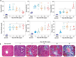
Plasma biochemical measurements indicated an increase in liver injury biomarkers and a dramatic increase in plasma bilirubin and bile acids at different time intervals after BDL surgery (Fig. 1A). Liver tissue histopathological alterations also revealed significant collagen deposition and fibrosis in this organ (Fig. 1B). These data could confirm the appropriate induction of cholestasis/cirrhosis in the current BDL model. The maximum collagen deposition and liver fibrosis were achieved on day 42 after BDL induction (Fig. 1B).

Fig. 1

Plasma biochemical analysis (A) and liver tissue histopathological alterations (B; blue colored, green arrow) in bile duct ligated (BDL) rats. These data confirm the occurrence of cholestasis in the current model. Data are shown as mean ±SD (n = 7). Data sets with various alphabetical superscripts differ significantly (p < 0.05). Scale bars for histopathological images are = 100 µm
/f/fulltexts/CEH/50386/CEH-9-50386-g001_min.jpg

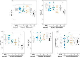
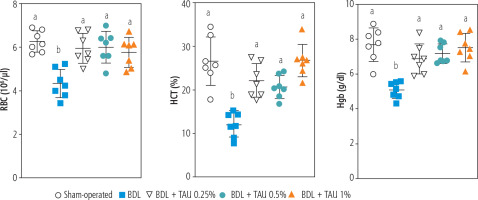
Blood analysis (CBC) of BDL rats revealed a significant decrease in erythrocyte count on day 42 after the BDL operation (Fig. 2). Other factors, such as Hgb and hematocrit (HTC), followed the same pattern and significantly decreased 42 days after BDL surgery (Fig. 2). Our model detected no significant changes in white blood cells (WBC) and platelet count at different intervals after BDL induction (Fig. 2). The effects of TAU on blood parameters were evaluated in BDL animals (Fig. 3). It was found that TAU (0.25%, 0.5%, and 1% w : v in drinking water for 42 consecutive days) significantly improved blood parameters in cirrhotic animals, including Hgb, HTC, and erythrocyte count. It should be mentioned that the effects of TAU on these parameters were not dose-dependent in the current model (Fig. 3).

Fig. 2

Complete blood count (CBC) and hematological alterations at different time points after bile duct ligated (BDL) surgery. A significant decrease in parameters associated with red blood cells (erythrocyte count, Hgb, and HCT) was detected in BDL rats 42 days after the BDL operation. No significant changes in WBC and platelets were detected in the current model. Data are presented as mean ±SD (n = 7). Data series with different alphabetical superscripts are significantly different (p < 0.05)
/f/fulltexts/CEH/50386/CEH-9-50386-g002_min.jpg
Fig. 3

Role of taurine (TAU) on complete blood count (CBC, RBC) of bile duct ligated (BDL) animals (42 days after BDL surgery). Data are represented as mean ±SD (n = 7). Data series with different alphabetical superscripts are significantly different (p < 0.05)
/f/fulltexts/CEH/50386/CEH-9-50386-g003_min.jpg

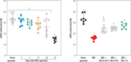
The activity of the enzyme G6PD in erythrocytes was another parameter assessed in the BDL animals. It was found that G6PD activity was significantly suppressed 42 days after the BDL operation. On the other hand, TAU (0.25%, 0.5%, and 1% w : v in drinking water for 42 consecutive days) significantly restored the G6PD activity in erythrocytes isolated from cirrhotic animals. No difference between various concentrations of TAU in improving G6PD activity was detected in the current study (Fig. 4).

Fig. 4

Monitoring glucose-6-deficiency (G6PD) at different intervals after bile duct ligation (BDL) surgery. A significant decrease in G6PD deficiency was detected 42 days after the BDL operation. It was found that taurine (TAU) significantly improved G6PD deficiency at both doses (0.25%, 0.5%, and 1% w : v). Data are shown as mean ±SD (n = 7). Data series with different alphabetical superscripts are significantly different (p < 0.05)
/f/fulltexts/CEH/50386/CEH-9-50386-g004_min.jpg

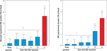
Morphological assessment of erythrocytes revealed the presence of hypochromic and polychromasia erythrocytes in the blood of BDL rats (42 days after BDL surgery; Fig. 5). No significant morphological changes in the blood of BDL rats were detected on days 3, 7, and 14 after the BDL operation (Fig. 5). It should be noted that TAU did not change erythrocyte morphological changes in the current model (data not shown).

Fig. 5

The light microscope analysis of erythrocytes in bile duct ligated (BDL) rats. A significant increase in microcytic hypochromic and polychromasia erythrocytes was detected in BDL rats. Taurine (TAU) had no significant effect on the RBS morphology in the current investigation. Data are shown as mean ±SD (n = 7). Data series with different alphabetical superscripts are significantly different (p < 0.05)
/f/fulltexts/CEH/50386/CEH-9-50386-g005_min.jpg

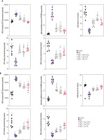
The assessment of biomarkers of oxidative stress at 28 and 42 days after BDL revealed significant ROS formation and lipid peroxidation in erythrocytes isolated from BDL rats. Moreover, the GSH level, CAT activity, and SOD activity of erythrocytes were significantly decreased in cirrhotic animals. It was found that TAU (0.25%, 0.5%, and 1%) significantly blunted markers of oxidative stress in erythrocytes of BDL rats. The effect of TAU on oxidative stress biomarkers was not dose-dependent in the current study (Fig. 6). As mentioned in various parts of the results, the effects of various doses of TAU (0.25%, 0.5%, and 1% w : v) on blood parameters assessed in the cirrhotic rats was not dose-dependent. Hence, the lower doses of this amino acid could be used for further investigations because of economic issues as well as preventing any potential adverse effects.

Fig. 6

Oxidative stress markers were assessed in erythrocytes on days 28 (A) and 42 after bile duct ligated (BDL) surgery (B). It was found that taurine (TAU; 0.5% and 1% w : v) significantly decreased biomarkers of oxidative stress in cirrhotic rats. In the current experiments, most oxidative stress markers were not significantly changed at 3, 7, and 14 days after cholestasis induction. Data are shown as mean ±SD (n = 7). Data series with different alphabetical superscripts are significantly different (p < 0.05)
/f/fulltexts/CEH/50386/CEH-9-50386-g006_min.jpg

Discussion

The cirrhosis-induced hematological complication is a severe clinical problem [11-16]. On the other hand, oxidative stress and its associated complications could play a significant role in this situation [11]. Several case reports of cholestasis/cirrhosis-induced anemia have been published [112-114]. Hypochromic microcytic anemia seems to be humans’ most prevalent phenotype of cholestasis/cirrhosis-induced anemia [112]. In the current study, we detected microcytic hypochromic RBCs in the BDL rats 42 days after the BDL operation (Fig. 5). This finding is in line with other studies that indicate microcytic anemia as a prevalent type of morphological change of RBCs in cirrhosis [112]. On the other hand, detecting a significant number of polychromasia erythrocytes was another interesting finding that was detected 28 and 42 days after BDL induction (Fig. 5). Polychromasia occurs when RBCs are prematurely released from the bone marrow to the blood stream [115]. We did not find any relevant study on the etiology of polychromasia in the BDL animal model of cirrhosis. Nevertheless, it has been reported that polychromasia is frequently detected in patients with chronic hepatitis or hepatocellular carcinoma [115]. The mechanism(s) of releasing such erythrocytes in the current model is ambiguous and warrants further studies. Low hemoglobin levels (e.g., 6 g/dl) have also been reported in cholestatic patients with anemia [112]. As mentioned, oxidative stress and its associated complications play an essential role in erythrocyte damage in cirrhosis. Therefore, it is important to find safe and clinically applicable agents for preventing erythrocyte disruption. The data obtained from this study revealed that TAU, as a safe amino acid with exciting features such as membrane stabilization, antioxidant action, and osmoregulatory properties, significantly protected erythrocytes in cirrhotic rats.

Cirrhosis-induced erythrocyte damage and lysis are widely investigated. Several mechanisms have been proposed to be involved in this complication. In this regard, oxidative stress and its associated complications in erythrocytes have received much attention [116-118]. Erythrocytes continuously transport a high concentration of oxygen over their lifespan. Therefore, it is well known that these cells are susceptible to exogenous and endogenous reactive species and oxidative stress [119]. However, different effective antioxidant systems have been developed during the evolution of these cells to counteract oxidative stress in erythrocytes [120, 121]. This system is mainly involved in the presence of an enzyme known as glucose-6-phosphate dehydrogenase (G6PD) (Fig. 7) [122, 123]. This enzyme reduces oxidized NADP+ to its reduced form (NADPH). The enzyme glutathione reductase uses NADPH to convert oxidized glutathione (GSSG) to its reduced form (Fig. 7). GSH and its associated enzymes are vital for counteracting oxidative stress in erythrocytes [124, 125]. GSH is widely used in erythrocytes to detoxify reactive species directly or as a substrate for GSH-dependent antioxidant enzymes (Fig. 7) [124, 125]. Many experimental models also mentioned decreased erythrocyte antioxidant levels (CAT, SOD, glutathione peroxidase [GPx]) [121, 126]. It is well known that SOD is the most crucial antioxidant in erythrocytes [127]. Superoxide anion (O2•-) is the most dangerous reactive species that damages erythrocytes and causes deformability [127].

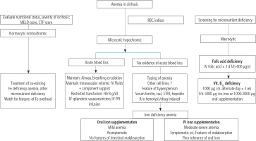
Fig. 7

Several options have been developed for managing anemia in cirrhosis in clinical settings. This figure is adapted from reference [17] (Creative Commons Attribution-NonCommercial; CC BY-NC 4.0); d – day, f – folic acid, b – bolus dose
/f/fulltexts/CEH/50386/CEH-9-50386-g007_min.jpg

Another critical issue regarding erythrocytes is their membrane disintegrability [128, 129]. Several mechanisms have been proposed in experimental models and human cases of this disease. For example, Verkleij et al. found that the fusion of abnormal plasma lipoproteins could lead to abnormal and enlarged RBCs in cholestatic patients [129]. In another study, Okano et al. found that lecithin and fatty acid composition of erythrocyte membrane are changed in biliary obstruction [128]. All these events could destabilize the erythrocyte membrane, leading to its deformities and rupture (Fig. 7). An exciting feature of TAU is its effect on cell membranes. It is well known that TAU is a membrane stabilizer [130]. The membrane stabilizing action of TAU on erythrocytes could render their membrane more resistant to reactive species such as ROS and lipid peroxidation byproducts [131-133]. Gossai et al. also found that TAU significantly stabilized the erythrocyte membrane and enhanced cellular antioxidant capacity [134]. Excitingly, Gossai et al. also found that TAU effectively prevents Hgb and lactate dehydrogenase (LDH) release from erythrocytes [134]. They administered TAU (300 mg/kg) to diabetic rats. The formation of ROS, oxidized glutathione, and malondialdehyde was significantly decreased in the erythrocytes of TAU-treated mice [134]. Moreover, levels of catalase, glutathione peroxidase, and superoxide dismutase were considerably higher in the RBC of diabetic erythrocytes [134]. In another study by Gossai et al., the turnover of GSSG to GSH in erythrocytes reduced oxidative stress [134].

An important mechanism proposed for erythrocyte death and anemia in cholestasis/cirrhosis is proposed by Lang et al. [135]. They investigated the pathogenic effect of bilirubin on erythrocytes. In the current investigation, we found that the level of plasma bilirubin was very high in BDL rats (> 10 mg/dl) (Fig. 1). Lang et al. found that mechanistically bilirubin caused a severe influx of Ca2+ to erythrocytes, accumulation of sphingomyelinase activation, formation of ceramide, and translocation of phosphatidyl serine to the erythrocyte surface [136]. They concluded that all these events could lead to erythrocyte death and anemia in cholestasis/cirrhosis. Interestingly, the protective effects of TAU against Ca2+ effects on many cells is an essential feature of this amino acid [136]. Hence this characteristic of TAU could play an essential role in its protective effect on erythrocytes of cirrhotic animals in the current study.

As mentioned, RBCs are continuously exposed to exogenous and endogenous oxidants with different etiologies [137]. Oxidative stress could cause a decrease in the erythrocyte’s life span, hemolysis, and, finally, erythrocyte death. In this regard, a plethora of investigations have studied the effects of antioxidant molecules against this complication. Several studies have mentioned the protective properties of antioxidant molecules such as β-carotene, polyphenols (e.g., resveratrol and quercetin), methionine, α-lipoic acid, N-acetylcysteine, melatonin, dithiothreitol (DTT), and homocysteine on erythrocytes damage with different etiologies [138-144]. Based on these data, using these molecules could significantly protect erythrocytes in various pathological conditions. Several options have been developed for managing anemia in cirrhosis in clinical settings (Fig. 7) [17]. Based on the study by Singh et al. [17], complementary treatment strategies could play a role in alleviating cirrhosis-induced anemia. First, removing the background diseases such as hepatitis or alcoholism might be very helpful [17]. Second, dietary advice (e.g., administration of elemental iron and other micronutrients) could help alleviate cirrhosis-induced anemia (Fig. 7). On the other hand, several studies have revealed the exciting effects of TAU on erythrocytes [38-43]. TAU is not considered a classic radical scavenger or a regulator of cellular antioxidant defense mechanisms [145-147]. Actually, TAU acts as a regulator of cellular ROS generation [145, 146, 148]. There is a plethora of evidence indicating the role of TAU in regulating mitochondria-mediated ROS formation [145, 146]. However, this mechanism is irrelevant to cells devoid of mitochondria (e.g., erythrocytes). Hence, another mechanism should be involved in the protective properties of TAU in erythrocytes. Interestingly, it has been proposed that TAU could have a regulatory effect on mitochondrial defense mechanisms or even the expression of the action of oxidants on mitochondrial-encoded proteins [149]. Then, the direct regulatory properties of TAU and its effects on cellular antioxidant mechanisms could play a pivotal role in its protective properties in biological systems.

Interestingly, the effect of TAU on erythrocytes’ G6PD enzyme activity has also been reported in previous studies [150, 151]. It was found that TAU significantly prevents suppression of the G6PD enzyme activity in erythrocytes exposed to toxic levels of cadmium, hydrogen peroxide, phenylhydrazine, and lead. In some studies it has been found that TAU could significantly increase the ratio of GSH/GSSG, which could directly increase the direct radical scavenging activity of GSH or indirectly enhance the activity of GSH-dependent enzyme systems (Fig. 7). Moreover, isolated erythrocytes have been challenged with H2O2 as a potent antioxidant [150, 151].The researchers also detected that TAU significantly prevented erythrocyte ATP breakdown, promoted GSH levels, and enhanced the pentode phosphate pathway [150, 151]. In the current investigation, we found that the oxidative stress markers were significantly elevated in cirrhotic rats. Another exciting finding of our study was the positive effects of TAU on the G6PD enzyme activity in erythrocytes of cirrhotic animals (Figs. 4 and 8). The higher activity of G6PD boosts erythrocytes’ antioxidant system and prevents cell death (Figs. 7 and 8). We also found that TAU significantly improved the antioxidant activity of erythrocytes isolated from cirrhotic animals (Fig. 6).

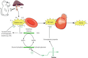
Fig. 8

Oxidative stress and its associated complications play a critical role in the pathogenesis of cirrhosis-induced erythrocyte damage, lysis, and anemia. The accumulation of potentially cytotoxic molecules, such as hydrophobic bile acids and supraphysiological concentrations of bilirubin, could be involved in the pathogenesis of these adverse effects
/f/fulltexts/CEH/50386/CEH-9-50386-g008_min.jpg

Another critical issue in erythrocyte storage and, finally, transfusion to recipients is the oxidation of the erythrocyte membrane. The oxidation of fatty acids ultimately compromises the stability of the erythrocytes’ membrane and eventually impairs their activity [152-154]. Interestingly, TAU is an excellent membrane stabilizer [130, 149, 155-157]. It has repeatedly been mentioned that TAU protects biomembrane lipids from oxidation and consequently prevents cell damage [130, 149, 155-157]. Huxtable et al. mentioned that inhibiting lipid peroxidation, stabilization of biological membranes, TAU antioxidant properties, and maintaining intracellular ions’ homeostasis play a crucial role in preserving cellular integrity and preventing their damage [23]. In the current study, we found that TAU treatment significantly mitigated hemolysis and lipid peroxidation in erythrocytes of cirrhotic animals (Fig. 6). Hence, this exciting feature of TAU could effectively prevent erythrocyte damage and hemolysis and play a crucial role in its protective properties in cirrhosis.

Treatments for cholestasis/cirrhosis-induced erythrocyte damage and anemia are symptomatic, and there are no specific pharmacological interventions to prevent or treat this complication. Hence, the data from this study might pave the way for finding novel and clinically applicable therapeutic options for preventing cholestasis/cirrhosis-induced erythrocyte damage and anemia (Fig. 8). Further studies are warranted to explain the precise mechanisms of TAU’s protective properties against erythrocyte damage and its application in clinical settings in cirrhotic patients, blood transfusion, and/or blood diseases with susceptible erythrocytes.

Acknowledgments

The authors acknowledge the Pharmaceutical Sciences Research Center for providing technical facilities for this investigation. This study was financially supported by the Vice-Chancellor of Research Affairs of Shiraz University of Medical Sciences (Grants: 23701/23031/23040/23028/16428/23024/23158/13555) and the Natural Science Foundation of Shanxi (Grant No. 20210302124411).

Disclosure

The authors declare no conflict of interest.

References

1 

Nusrat S, Khan MS, Fazili J, Madhoun MF. Cirrhosis and its complications: Evidence based treatment. World J Gastroenterol 2014; 20: 5442-5460.

2 

Heidari R, Moezi L, Asadi B, et al. Hepatoprotective effect of boldine in a bile duct ligated rat model of cholestasis/cirrhosis. Pharma Nutrition 2017; 5: 109-117.

3 

Abdoli N, Sadeghian I, Mousavi K, et al. Suppression of cirrhosis related renal injury by N-acetyl cysteine. Curr Res Pharmacol Drug Discov 2020; 1: 30-38.

4 

Ommati MM, Farshad O, Niknahad H, et al. Cholestasis-associated reproductive toxicity in male and female rats: The fundamental role of mitochondrial impairment and oxidative stress. Toxicol Lett 2019; 316: 60-72.

5 

Ommati MM, Mohammadi H, Mousavi K, et al. Metformin alleviates cholestasis-associated nephropathy through regulating oxidative stress and mitochondrial function. Liver Res 2021; 5: 171-180.

6 

Ghanbarinejad V, Jamshidzadeh A, Khalvati B, et al. Apoptosisinducing factor plays a role in the pathogenesis of hepatic and renal injury during cholestasis. Naunyn Schmiedebergs Arch Pharmacol 2021; 394: 1191-1203.

7 

Ommati MM, Hojatnezhad S, Abdoli N, et al. Pentoxifylline mitigates cholestasis-related cholemic nephropathy. Clin Exp Hepatol 2021; 7: 377-389.

8 

Ommati MM, Farshad O, Azarpira N, et al. Silymarin mitigates bile duct obstruction-induced cholemic nephropathy. Naunyn Schmiedebergs Arch Pharmacol 2021; 394: 1301-1314.

9 

Jamshidzadeh A, Heidari R, Latifpour Z, et al. Carnosine ameliorates liver fibrosis and hyperammonemia in cirrhotic rats. Clin Res Hepatol Gastroenterol 2017; 41: 424-434.

10 

Heidari R. Brain mitochondria as potential therapeutic targets for managing hepatic encephalopathy. Life Sci 2019; 218: 65-80.

11 

Grattagliano I, Calamita G, Cocco T, et al. Pathogenic role of oxidative and nitrosative stress in primary biliary cirrhosis. World J Gastroenterol 2014; 20: 5746-5759.

12 

Muriel P, Favari L, Soto C. Erythrocyte alterations correlate with CCl4 and biliary obstruction-induced liver damage in the rat. Life Sci 1993; 52: 647-655.

13 

Masako T, Fumihiko T, Hideto I, Toshio S. Experimental biliary obstruction of rat: Initial changes in the structure and lipid content of erythrocytes. Biochim Biophys Acta 1983; 753: 22-31.

14 

Heuman DM, Pandak WM, Hylemon PB, Vlahcevic ZR. Conjugates of ursodeoxycholate protect against cytotoxicity of more hydrophobic bile salts: In vitro studies in rat hepatocytes and human erythrocytes. Hepatology 1991; 14: 920-926.

15 

Premkumar M. Assessment of role of iron metabolism, nutritional deficiency, gastrointestinal loss, and chronic inflammation in the etiopathogenesis of anemia in chronic liver disease. Clinicaltrials.gov, 2021/09/26/2021. NCT04622449.

16 

Manrai M, Dawra S, Kapoor R, et al. Anemia in cirrhosis: An underestimated entity. World J Clin Case 2022; 10: 777-789.

17 

Singh A, Srivastava S, Kapoor R, et al. Anemia in cirrhosis: An underestimated entity. World J Clin Cases 2022; 10: 777-789.

18 

Geetha A, Lakshmi Priya MD, Jeyachristy SA, Surendran R. Level of oxidative stress in the red blood cells of patients with liver cirrhosis. Indian J Med Res 2007; 126: 204-210.

19 

Ripps H, Shen W. Review: Taurine: A “very essential” amino acid. Mol Vis 2012; 18: 2673-2686.

20 

Jacobsen JG, Smith LH. Biochemistry and physiology of taurine and taurine derivatives. Physiol Rev 1968; 48: 424-511.

21 

Bouckenooghe T, Remacle C, Reusens B. Is taurine a functional nutrient? Curr Opin Clin Nutr Metab Care 2006; 9: 728-733.

22 

Huxtable RJ, Michalk D. Taurine in health and disease. Springer Science & Business Media, New York 2013.

23 

Huxtable RJ. Physiological actions of taurine. Physiol Rev 1992; 72: 101-163.

24 

De Carvalho FG, Galan BSM, Santos PC, et al. Taurine: A potential ergogenic aid for preventing muscle damage and protein catabolism and decreasing oxidative stress produced by endurance exercise. Front Physiol 2017; 8: 710.

25 

Han X, Chesney RW. The role of taurine in renal disorders. Amino Acids 2012; 43: 2249-2263.

26 

Hansen SH, Grunnet N. Taurine, glutathione and bioenergetics. In: Idrissi AE, L’Amoreaux WJ (Eds.). Taurine 8. Springer, New York 2013: 3-12.

27 

Rodella P, Takase L, Santos J, et al. The effect of taurine on hepatic disorders. Curr Updates Hepatol Gastroenterol 2017; 1: 1-12.

28 

Najibi A, Rezaei H, Manthari RK, et al. Cellular and mitochondrial taurine depletion in bile duct ligated rats: a justification for taurine supplementation in cholestasis/cirrhosis. Clin Exp Hepatol 2022; 8: 195-210.

29 

Hansen SH, Andersen ML, Birkedal H, et al. The important role of taurine in oxidative metabolism. In: Oja SS, Saransaari P (Eds.). Taurine 6. Springer, US 2006; 129-135.

30 

Jamshidzadeh A, Heidari R, Abasvali M, et al. Taurine treatment preserves brain and liver mitochondrial function in a rat model of fulminant hepatic failure and hyperammonemia. Biomed Pharmacother 2017; 86: 514-520.

31 

Heidari R, Babaei H, Eghbal MA. Amodiaquine-induced toxicity in isolated rat hepatocytes and the cytoprotective effects of taurine and/or N-acetyl cysteine. Res Pharm Sci 2014; 9: 97-105.

32 

Heidari R, Babaei H, Eghbal MA. Cytoprotective effects of taurine against toxicity induced by isoniazid and hydrazine in isolated rat hepatocytes. Arh Hig Rada Toksikol 2013; 64: 201-209.

33 

Heidari R, Babaei H, Eghbal MA. Ameliorative effects of taurine against methimazole-induced cytotoxicity in isolated rat hepatocytes. Sci Pharm 2012; 80: 987-1000.

34 

Niknahad H, Jamshidzadeh A, Heidari R, et al. Ammonia induced mitochondrial dysfunction and energy metabolism disturbances in isolated brain and liver mitochondria, and the effect of taurine administration: relevance to hepatic encephalopathy treatment. Clin Exp Hepatol 2017; 3: 141-151.

35 

Jamshidzadeh A, Niknahad H, Heidari R, et al. Carnosine protects brain mitochondria under hyperammonemic conditions: Relevance to hepatic encephalopathy treatment. Pharma Nutrition 2017; 5: 58-63.

36 

Ommati MM, Heidari R, Ghanbarinejad V, et al. Taurine treatment provides neuroprotection in a mouse model of manganism. Biol Trace Elem Res 2019; 190: 384-395.

37 

Ahmadi N, Ghanbarinejad V, Ommati MM, et al. Taurine prevents mitochondrial membrane permeabilization and swelling upon interaction with manganese: Implication in the treatment of cirrhosis-associated central nervous system complications. J Biochem Mol Toxicol 2018; 32: e22216.

38 

Bertolone L, Roy MK, Hay AM, et al. Impact of taurine on red blood cell metabolism and implications for blood storage. Transfusion 2020; 60: 1212-1226.

39 

Pokhrel PK, Lau-Cam CA. Protection by taurine and structurally related sulfur-containing compounds against erythrocyte membrane damage by hydrogen peroxide. In: Della Corte L, Huxtable RJ, Sgaragli G, Tipton KF (Eds.). Taurine 4: Taurine and Excitable Tissues. Boston, MA, Springer US 2002; 411-429.

40 

Sinha M, Manna P, Sil PC. Taurine protects the antioxidant defense system in the erythrocytes of cadmium treated mice. BMB Rep 2008; 41: 657-663.

41 

Ali SN, Arif A, Ansari FA, Mahmood R. Cytoprotective effect of taurine against sodium chlorate-induced oxidative damage in human red blood cells: an ex vivo study. Amino Acids 2022; 54: 33-46.

42 

Ansari FA, Ali SN, Mahmood R. Taurine mitigates nitriteinduced methemoglobin formation and oxidative damage in human erythrocytes. Environ Sci Pollut Res Int 2017; 24: 19086-19097.

43 

Husain N, Mahmood R. Taurine attenuates Cr(VI)-induced cellular and DNA damage: an in vitro study using human erythrocytes and lymphocytes. Amino Acids 2020; 52: 35-53.

44 

Heidari R, Abdoli N, Ommati MM, et al. Mitochondrial impairment induced by chenodeoxycholic acid: The protective effect of taurine and carnosine supplementation. Trend Pharm Sci 2018; 4.

45 

Jamshidzadeh A, Abdoli N, Niknahad H, et al. Taurine alleviates brain tissue markers of oxidative stress in a rat model of hepatic encephalopathy. Trend Pharm Sci 2017; 3: 181-192.

46 

Heidari R, Jamshidzadeh A, Niknahad H, et al. The hepatoprotection provided by taurine and glycine against antineoplastic drugs induced liver injury in an ex vivo model of normothermic recirculating isolated perfused rat liver. Trend Pharm Sci 2016; 2: 59-76.

47 

Mousavi K, Niknahad H, Ghalamfarsa A, et al. Taurine mitigates cirrhosis-associated heart injury through mitochondrial-dependent and antioxidative mechanisms. Clin Exp Hepatol 2020; 6: 207-219.

48 

Farshad O, Ommati MM, uuml, et al. Skeletal muscle mitochondrial impairment in cirrhosis-induced sarcopenia. Trend Pharm Sci 2020; 6: 189-204.

49 

Ommati MM, Farshad O, Jamshidzadeh A, Heidari R. Taurine enhances skeletal muscle mitochondrial function in a rat model of resistance training. Pharma Nutrition 2019; 9: 100161.

50 

Ommati MM, Mobasheri A, Ma Y, et al. Taurine mitigates the development of pulmonary inflammation, oxidative stress, and histopathological alterations in a rat model of bile duct ligation. Naunyn Schmiedebergs Arch Pharmacol 2022; 395: 1557-1572.

51 

Heidari R, Jamshidzadeh A, Ghanbarinejad V, et al. Taurine supplementation abates cirrhosis-associated locomotor dysfunction. Clin Exp Hepatol 2018; 4: 72-82.

52 

Moezi L, Heidari R, Amirghofran Z, et al. Enhanced anti-ulcer effect of pioglitazone on gastric ulcers in cirrhotic rats: The role of nitric oxide and IL-1β. Pharmacol Rep 2013; 65: 134-143.

53 

Ommati MM, Farshad O, Azarpira N, et al. Betaine alleviates cholestasis-associated renal injury by mitigating oxidative stress and enhancing mitochondrial function. Biologia 2021; 76: 351-365.

54 

Siavashpour A, Khalvati B, Azarpira N, et al. Poly (ADP-Ribose) polymerase-1 (PARP-1) overactivity plays a pathogenic role in bile acids-induced nephrotoxicity in cholestatic rats. Toxicol Lett 2020; 330: 144-158.

55 

Ahmadi A, Niknahad H, Li H, et al. The inhibition of NF-кB signaling and inflammatory response as a strategy for blunting bile acid-induced hepatic and renal toxicity. Toxicol Lett 2021; 349: 12-29.

56 

Ommati MM, Amjadinia A, Mousavi K, et al. N-acetyl cysteine treatment mitigates biomarkers of oxidative stress in different tissues of bile duct ligated rats. Stress 2021; 24: 213-228.

57 

Mousavi K, Niknahad H, Li H, et al. The activation of nuclear factor-E2-related factor 2 (Nrf2)/heme oxygenase-1 (HO-1) signaling blunts cholestasis-induced liver and kidney injury. Toxicol Res 2021; 10: 911-927.

58 

Ommati MM, Farshad O, Mousavi K, et al. Agmatine alleviates hepatic and renal injury in a rat model of obstructive jaundice. Pharma Nutrition 2020; 13: 100212.

59 

Ommati MM, Farshad O, Niknahad H, et al. Oral administration of thiol-reducing agents mitigates gut barrier disintegrity and bacterial lipopolysaccharide translocation in a rat model of biliary obstruction. Curr Res Pharmacol Drug Discov 2020; 1: 10-18.

60 

Heidari R, Mohammadi H, Ghanbarinejad V, et al. Proline supplementation mitigates the early stage of liver injury in bile duct ligated rats. J Basic Clin Physiol Pharmacol 2019; 30: 91-101.

61 

Ommati MM, Farshad O, Mousavi K, et al. Betaine supplementation mitigates intestinal damage and decreases serum bacterial endotoxin in cirrhotic rats. Pharma Nutrition 2020; 12: 100179.

62 

Heidari R, Niknahad H, Sadeghi A, et al. Betaine treatment protects liver through regulating mitochondrial function and counteracting oxidative stress in acute and chronic animal models of hepatic injury. Biomed Pharmacother 2018; 103: 75-86.

63 

Heidari R, Niknahad H. The role and study of mitochondrial impairment and oxidative stress in cholestasis. Methods Mol Biol 2019; 1981: 117-132.

64 

Mosleh G, Azadi A, Khademian S, et al. Anti-inflammatory activity and quality control of Erysimum cheiri (L.) Crantz.Biomed Res Int 2021; 2021: e5526644.

65 

Heidari R, Jafari F, Khodaei F, et al. Mechanism of valproic acid-induced Fanconi syndrome involves mitochondrial dysfunction and oxidative stress in rat kidney. Nephrology 2018; 23: 351-361.

66 

Ommati MM, Heidari R, Jamshidzadeh A, et al. Dual effects of sulfasalazine on rat sperm characteristics, spermatogenesis, and steroidogenesis in two experimental models. Toxicol Lett 2018; 284: 46-55.

67 

Ommati MM, Farshad O, Mousavi K, et al. Chlorogenic acid supplementation improves skeletal muscle mitochondrial function in a rat model of resistance training. Biologia 2020; 75: 1221-1230.

68 

Heidari R, Ghanbarinejad V, Mohammadi H, et al. Dithiothreitol supplementation mitigates hepatic and renal injury in bile duct ligated mice: Potential application in the treatment of cholestasis-associated complications. Biomed Pharmacother 2018; 99: 1022-1032.

69 

Heidari R, Mandegani L, Ghanbarinejad V, et al. Mitochondrial dysfunction as a mechanism involved in the pathogenesis of cirrhosis-associated cholemic nephropathy. Biomed Pharmacother 2019; 109: 271-280.

70 

Abdoli N, Sadeghian I, Azarpira N, et al. Taurine mitigates bile duct obstruction-associated cholemic nephropathy: effect on oxidative stress and mitochondrial parameters. Clin Exp Hepatol 2021; 7: 30-40.

71 

Hermann PB, Pianovski MAD, Henneberg R, et al. Erythrocyte oxidative stress markers in children with sickle cell disease. J Pediatr (Rio J) 2016; 92: 394-399.

72 

Ommati MM, Attari H, Siavashpour A, et al. Mitigation of cholestasis-associated hepatic and renal injury by edaravone treatment: Evaluation of its effects on oxidative stress and mitochondrial function. Liver Res 2021; 5: 181-193.

73 

Ommati MM, Heidari R, Zamiri MJ, et al. The footprints of oxidative stress and mitochondrial impairment in arsenic trioxide-induced testosterone release suppression in pubertal and mature F1-male Balb/c mice via the downregulation of 3β-HSD, 17β-HSD, and CYP11a expression. Biol Trace Elem Res 2020; 195: 125-134.

74 

Akram J, Reza H, Farzaneh A, et al. Antimalarial drugs-induced hepatic injury in rats and the protective role of carnosine. Pharm Sci 2016; 22: 170-180.

75 

Abdoli N, Heidari R, Azarmi Y, Eghbal MA. Mechanisms of the statins cytotoxicity in freshly isolated rat hepatocytes. J Biochem Mol Toxicol 2013; 27: 287-294.

76 

Heidari R, Behnamrad S, Khodami Z, et al. The nephroprotective properties of taurine in colistin-treated mice is mediated through the regulation of mitochondrial function and mitigation of oxidative stress. Biomed Pharmacother 2019; 109: 103-111.

77 

Heidari R, Arabnezhad MR, Ommati MM, et al. Boldine supplementation regulates mitochondrial function and oxidative stress in a rat model of hepatotoxicity. Pharm Sci 2019; 25: 1-10.

78 

Ommati MM, Shi X, Li H, et al. The mechanisms of arsenic-induced ovotoxicity, ultrastructural alterations, and autophagic related paths: An enduring developmental study in folliculogenesis of mice. Ecotoxicol Environ Saf 2020; 204: 110973.

79 

Ommati MM, Heidari R, Ghanbarinejad V, et al. The neuroprotective properties of carnosine in a mouse model of manganism is mediated via mitochondria regulating and antioxidative mechanisms. Nutr Neurosci 2020; 23: 731-743.

80 

Ommati MM, Manthari RK, Tikka C, et al. Arsenic-induced autophagic alterations and mitochondrial impairments in HPG-S axis of mature male mice offspring (F1-generation): A persistent toxicity study. Toxicol Lett 2020; 326: 83-98.

81 

Emadi E, Abdoli N, Ghanbarinejad V, et al. The potential role of mitochondrial impairment in the pathogenesis of imatinib-induced renal injury. Heliyon 2019; 5: e01996.

82 

Mobasher P, Ommati MM, Najibi A, et al. Searching for alternative toxicology testing systems: The response of isolated mitochondria from Saccharomyces cerevisiae, potato tuber, and mouse liver to a toxic insult. Trends Pharm Sci 2021; 7: 179-190.

83 

Pasdaran A, Azarpira N, Heidari R, et al. Effects of some cosmetic dyes and pigments on the proliferation of human foreskin fibroblasts and cellular oxidative stress; potential cytotoxicity of chlorophyllin and indigo carmine on fibroblasts. J Cosmet Dermatol 2022; 21: 3979-3985.

84 

Shafiekhani M, Ommati MM, Azarpira N, et al. Glycine supplementation mitigates lead-induced renal injury in mice. J Exp Pharmacol 2019; 11: 15-22.

85 

Ommati MM, Jamshidzadeh A, Heidari R, et al. Carnosine and histidine SsLead-induced reproductive toxicity through antioxidative and mitochondria-dependent mechanisms. Biol Trace Elem Res 2019; 187: 151-162.

86 

Heidari R, Ahmadi A, Mohammadi H, et al. Mitochondrial dysfunction and oxidative stress are involved in the mechanism of methotrexate-induced renal injury and electrolytes imbalance. Biomed Pharmacother 2018; 107: 834-840.

87 

Farshad O, Heidari R, Zamiri MJ, et al. Spermatotoxic effects of single-walled and multi-walled carbon nanotubes on male mice. Front Vet Sci 2020; 7: 591558.

88 

Eftekhari A, Ahmadian E, Azarmi Y, et al. In vitro/vivo studies towards mechanisms of risperidone-induced oxidative stress and the protective role of coenzyme Q10 and N-acetylcysteine. Toxicol Mech Methods 2016; 26: 520-528.

89 

Ommati MM, Arabnezhad MR, Farshad O, et al. The role of mitochondrial impairment and oxidative stress in the pathogenesis of lithium-induced reproductive toxicity in male mice. Front Vet Sci 2021; 8: 603262.

90 

Ommati MM, Jamshidzadeh A, Heidari R, et al. Carnosine and histidine supplementation blunt lead-induced reproductive toxicity through antioxidative and mitochondria-dependent mechanisms. Biol Trace Elem Res 2019; 187: 151-162.

91 

Heidari R, Babaei H, Eghbal M. Mechanisms of methimazole cytotoxicity in isolated rat hepatocytes. Drug Chem Toxicol 2013; 36: 403-411.

92 

Heidari R, Niknahad H, Jamshidzadeh A, et al. Carbonyl traps as potential protective agents against methimazole-induced liver injury. J Biochem Mol Toxicol 2015; 29: 173-181.

93 

Heidari R, Taheri V, Rahimi HR, et al. Sulfasalazine-induced renal injury in rats and the protective role of thiol-reductants. Ren Fail 2016; 38: 137-141.

94 

Jamshidzadeh A, Heidari R, Razmjou M, et al. An in vivo and in vitro investigation on hepatoprotective effects of Pimpinella anisum seed essential oil and extracts against carbon tetrachloride-induced toxicity. Iran J Basic Med Sci 2015; 18: 205-211.

95 

Heidari R, Esmailie N, Azarpira N, et al. Effect of thiol-reducing agents and antioxidants on sulfasalazine-induced hepatic injury in normotermic recirculating isolated perfused rat liver. Toxicol Res 2016; 32: 133-140.

96 

Heidari R, Babaei H, Eghbal MA. Cytoprotective effects of organosulfur compounds against methimazole induced toxicity in isolated rat hepatocytes. Adv Pharm Bull 2013; 3: 135-142.

97 

Heidari R, Jamshidzadeh A, Niknahad H, et al. Effect of taurine on chronic and acute liver injury: Focus on blood and brain ammonia. Toxicol Rep 2016; 3: 870-879.

98 

Duranti G, Ceci R, Patrizio F, et al. Chronic consumption of quercetin reduces erythrocytes oxidative damage: Evaluation at resting and after eccentric exercise in humans. Nutr Res 2018; 50: 73-81.

99 

Niknahad H, Heidari R, Alzuhairi AM, Najibi A. Mitochondrial dysfunction as a mechanism for pioglitazone-induced injury toward HepG2 cell line. Pharm Sci 2015; 20: 169-174.

100 

Ommati MM, Jamshidzadeh A, Niknahad H, et al. N-acetylcysteine treatment blunts liver failure-associated impairment of locomotor activity. Pharma Nutrition 2017; 5: 141-147.

101 

Ommati MM, Heidari R, Manthari RK, et al. Paternal exposure to arsenic resulted in oxidative stress, autophagy, and mitochondrial impairments in the HPG axis of pubertal male offspring. Chemosphere 2019; 236: 124325.

102 

Jamshidzadeh A, Niknahad H, Heidari R, et al. Propylthiouracil-induced mitochondrial dysfunction in liver and its relevance to drug-induced hepatotoxicity. Pharm Sci 2017; 23: 95-102.

103 

Ommati MM, Ahmadi HN, Sabouri S, et al. Glycine protects the male reproductive system against lead toxicity via alleviating oxidative stress, preventing sperm mitochondrial impairment, improving kinematics of sperm, and blunting the downregulation of enzymes involved in the steroidogenesis. Environ Toxicol 2022; 37: 2990-3006.

104 

Ommati MM, Abdoli N, Firoozi M, et al. Sildenafil blunts lung inflammation and oxidative stress in a rat model of cholestasis. Pharm Sci 2022 (in press).

105 

Ahmadian E, Babaei H, Nayebi AM, et al. Mechanistic approach for toxic effects of bupropion in primary rat hepatocytes. Drug Res 2017; 67: 217-222.

106 

Ommati MM, Li H, Jamshidzadeh A, et al. The crucial role of oxidative stress in non-alcoholic fatty liver disease-induced male reproductive toxicity: the ameliorative effects of Iranian indigenous probiotics. Naunyn Schmiedebergs Arch Pharmacol 2022; 395: 247-265.

107 

Ahmadian E, Eftekhari A, Fard JK, et al. In vitro and in vivo evaluation of the mechanisms of citalopram-induced hepatotoxicity. Arch Pharm Res 2017; 40: 1296-1313.

108 

Farshad O, Heidari R, Mohammadi H, et al. Hepatoprotective effects of Avicennia Marina (Forssk.) Vierh.Trends Pharm Sci 2017; 3: 255-266.

109 

Jamshidzadeh A, Dabagh F, Farshad O, et al. Hepatoprotective properties of the glycolipoprotein extract from Eisenia foetida. Trends Pharm Sci 2018; 4: 149-160.

110 

Jamshidzadeh A, Heidari R, Golzar T, Derakhshanfar A. Effect of Eisenia foetida extract against cisplatin-induced kidney injury in rats. J Diet Suppl 2016; 13: 551-559.

111 

Heidari R, Babaei H, Roshangar L, Eghbal MA. Effects of enzyme induction and/or glutathione depletion on methimazole-induced hepatotoxicity in mice and the protective role of N-acetylcysteine. Adv Pharm Bull 2014; 4: 21-28.

112 

Law ST, Lee WK, Li MKK, Lok KH. A gentleman with anemia and cholestasis. Case Report Med 2010; 2010: 536207.

113 

Altintaş E, Tiftik EN, Uçbilek E, Sezgin O. Sickle cell anemia connected with chronic intrahepatic cholestasis: a case report. Turk J Gastroenterol 2003; 14: 215-218.

114 

Fiel MI, Schiano T. A woman with chronic anemia and cholestatic liver disease. Hepatology 2009; 49: 1390-1391.

115 

Ohshima S, Toyama K. Morphological abnormalities of red blood cells in liver diseases. Rinsho Ketsueki 1987; 28: 1717-1722.

116 

Morse EE. Mechanisms of hemolysis in liver disease. Ann Clin Lab Sci 1990; 20: 169-174.

117 

Mei C, Peng F, Yin W, et al. Increased suicidal erythrocyte death in patients with hepatitis B-related acute-on-chronic liver failure. Am J Physiol 2022; 323: G9-G20.

118 

Gonzalez-Casas R, Jones EA, Moreno-Otero R. Spectrum of anemia associated with chronic liver disease. World J Gastroenterol 2009; 15: 4653-4658.

119 

Edwards CJ, Fuller J. Oxidative stress in erythrocytes. Compar Haematol Int 1996; 6: 24-31.

120 

Kurata M, Suzuki M, Agar NS. Antioxidant systems and erythrocyte life-span in mammals. Comp Biochem Physiol 1993; 106: 477-487.

121 

Çimen MYB. Free radical metabolism in human erythrocytes. Clin Chim Acta 2008; 390: 1-11.

122 

Efferth T, Schwarzl SM, Smith J, Osieka R. Role of glucose-6-phosphate dehydrogenase for oxidative stress and apoptosis. Cell Death Differ 2006; 13: 527-528.

123 

Franco R, Navarro G, Martínez-Pinilla E. Antioxidant defense mechanisms in erythrocytes and in the central nervous system. Antioxidants 2019; 8: 46.

124 

Harley JD. Role of reduced glutathione in human erythrocytes. Nature 1965; 206: 1054-1055.

125 

Raftos JE, Whillier S, Kuchel PW. Glutathione synthesis and turnover in the human erythrocyte: Alignment of a model based on detailed enzyme kinetics with experimental data. J Biol Chem 2010; 285: 23557-23567.

126 

Doustimotlagh AH, Dehpour AR, Nourbakhsh M, Golestani A. Alteration in membrane protein, antioxidant status and hexokinase activity in erythrocytes of CCl4-induced cirrhotic rats. Acta Med Iran 2014; 52: 795-803.

127 

Uyesaka N, Hasegawa S, Ishioka N, et al. Effects of superoxide anions on red cell deformability and membrane proteins. Biorheology 1992; 29: 217-229.

128 

Okano Y, Yamauchi T, Sekiya T, et al. Mechanism for lipid abnormalities of erythrocyte membranes in biliary obstruction: lecithin content and its fatty acyl composition. Clin Chim Acta 1978; 88: 237-248.

129 

Verkleij AJ, Nauta IL, Werre JM, et al. The fusion of abnormal plasma lipoprotein (LP-X) and the erythrocyte membrane in patients with cholestasis studied by electronmicroscopy. Biochim Biophys Acta 1976; 436: 366-376.

130 

Schaffer SW, Allo S, Harada H, et al. Mechanism underlying the membrane-stabilizing activity of taurine. In: Iwata H, Lombardini JB, Segawa T (Eds.). Taurine and the Heart: Proceedings of the Symposium Annexed to the 10th Annual Meeting of the Japanese Research Society on Sulfur Amino Acids Osaka, Japan, September 10, 1987. Springer, Boston, MA 1989; 43-50.

131 

Schaffer S, Kim HW. Effects and mechanisms of taurine as a therapeutic agent. Biomol Ther (Seoul) 2018; 26: 225-241.

132 

Yayu W. Effect of combination of puerarin and taurine on stability of erythrocytes. Trad Chin Drug Res Clin Pharmacol 2014.

133 

Akhalaya MY, Kushnareva EA, Parshina EY, et al. Membrane-modifying effect of taurine. Biophysics 2012; 57: 485-490.

134 

Gossai D, Lau-Cam CA. The effects of taurine, taurine homologs and hypotaurine on cell and membrane antioxidative system alterations caused by type 2 diabetes in rat erythrocytes. Adv Exp Med Biol 2009; 643: 359-368.

135 

Lang E, Gatidis S, Freise NF, et al. Conjugated bilirubin triggers anemia by inducing erythrocyte death. Hepatology 2015; 61: 275-284.

136 

Lang E, Gatidis S, Freise NF, et al. Conjugated bilirubin triggers anemia by inducing erythrocyte death. Hepatology (Baltimore, Md.) 2015; 61: 275-284.

137 

Mohanty JG, Nagababu E, Rifkind JM. Red blood cell oxidative stress impairs oxygen delivery and induces red blood cell aging. Front Physiol 2014; 5: 84.

138 

Jamshidzadeh A, Rezaeian Mehrabadi A. Protective effect of quercetin on oxidative stress in glucose-6-phosphate dehydrogenase-deficient erythrocytes in vitro. Iran J Pharm Res 2010; 9: 169-175.

139 

Pandey KB, Rizvi SI. Protective effect of resveratrol on markers of oxidative stress in human erythrocytes subjected to in vitro oxidative insult. Phytother Res 2010; 24 Suppl 1: S11-14.

140 

Ghibu S, Lauzier B, Delemasure S, et al. Antioxidant properties of alpha-lipoic acid: effects on red blood membrane permeability and adaptation of isolated rat heart to reversible ischemia. Mol Cell Biochem 2009; 320: 141-148.

141 

Selvam R, Anuradha CV. Effect of oral methionine on blood lipid peroxidation and antioxidants in alloxan-induced diabetic rats. J Nutr Biochem 1990; 1: 653-658.

142 

Al Balushi H, Hannemann A, Rees D, et al. The effect of antioxidants on the properties of red blood cells from patients with sickle cell anemia. Front Physiol 2019; 10: 975.

143 

Caylak E, Aytekin M, Halifeoglu I. Antioxidant effects of methionine, α-lipoic acid, N-acetylcysteine and homocysteine on lead-induced oxidative stress to erythrocytes in rats. Exp Toxicol Pathol 2008; 60: 289-294.

144 

da Rosa DP, Bona S, Simonetto D, et al. Melatonin protects the liver and erythrocytes against oxidative stress in cirrhotic rats. Arq Gastroenterol 2010; 47: 72-78.

145 

Schaffer S, Azuma J, Takahashi K, Mozaffari M. Why is taurine cytoprotective? In: Lombardini JB, Schaffer SW, Azuma J (Eds.). Taurine 5: Beginning the 21st Century. Springer, Boston, MA 2003; 307-321.

146 

Hansen SH, Andersen ML, Cornett C, et al. A role for taurine in mitochondrial function. J Biomed Sci 2010; 17: S23.

147 

Heidari R, Jamshidzadeh A, Keshavarz N, Azarpira N. Mitigation of methimazole-induced hepatic injury by taurine in mice. Sci Pharm 2015; 83: 143-158.

148 

Heidari R, Rasti M, Shirazi Yeganeh B, et al. Sulfasalazine-induced renal and hepatic injury in rats and the protective role of taurine. Bioimpacts 2016; 6: 3-8.

149 

Zhang Z, Liu DAN, Yi BO, et al. Taurine supplementation reduces oxidative stress and protects the liver in an iron-overload murine model. Mol Med Report 2014; 10: 2255-2262.

150 

Gürer H, Ozgünes H, Saygin E, Ercal N. Antioxidant effect of taurine against lead-induced oxidative stress. Arch Environ Contam Toxicol 2001; 41: 397-402.

151 

Aruoma OI, Halliwell B, Hoey BM, Butler J. The antioxidant action of taurine, hypotaurine and their metabolic precursors. Biochem J 1988; 256: 251-255.

152 

Howie HL, Hay AM, de Wolski K, et al. Differences in Steap3 expression are a mechanism of genetic variation of RBC storage and oxidative damage in mice. Blood Adv 2019; 3: 2272-2285.

153 

de Wolski K, Fu X, Dumont LJ, et al. Metabolic pathways that correlate with post-transfusion circulation of stored murine red blood cells. Haematologica 2016; 101: 578-586.

154 

Hay A, Howie HL, Waterman HR, et al. Murine red blood cells from genetically distinct donors cross-regulate when stored together. Transfusion 2017; 57: 2657-2664.

155 

Huxtable R, Bressler R. Effect of taurine on a muscle intracellular membrane. Biochim Biophys Acta 1973; 323: 573-583.

156 

Sirdah MM, Abushahla AK, Al-Sarraj HAA. Effect of the addition of the antioxidant taurine on the complete blood count of whole blood stored at room temperature and at 4ºC for up to 7 days. Revista Brasil Hematol Hemoterap 2013; 35: 44-51.

157 

Schaffer SW, Ju Jong C, Kc R, Azuma J. Physiological roles of taurine in heart and muscle. J Biomed Sci 2010; 17: S2.

158 

Singh S, Manrai M, Parvathi VS, et al. Association of liver cirrhosis severity with anemia: does it matter? Ann Gastroenterol 2020; 33: 272-276.

159 

Özatli D, Köksal AS, Haznedaroglu IC, et al. Erythrocytes: anemias in chronic liver diseases. Hematology 2000; 5: 69-76.

160 

Gkamprela E, Deutsch M, Pectasides D. Iron deficiency anemia in chronic liver disease: etiopathogenesis, diagnosis and treatment. Ann Gastroenterol 2017; 30: 405-413.

161 

Selinger CP, Ang YS. Gastric antral vascular ectasia (GAVE): an update on clinical presentation, pathophysiology and treatment. Digestion 2008; 77: 131-137.

Copyright: © Clinical and Experimental Hepatology. This is an Open Access journal, all articles are distributed under the terms of the Creative Commons Attribution-NonCommercial-ShareAlike 4.0 International (CC BY-NC-SA 4.0). License (http://creativecommons.org/licenses/by-nc-sa/4.0/) enables reusers to distribute, remix, adapt, and build upon the material in any medium or format for noncommercial purposes only, and only so long as attribution is given to the creator. If you remix, adapt, or build upon the material, you must license the modified material under identical terms.
 
Quick links
© 2024 Termedia Sp. z o.o.
Developed by Bentus.