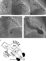
Herein, we present the case of a 54-year-old female who presented to the Emergency Department with central substernal chest pain lasting for 0.5 h prior to her arrival. Upon examination, she reported no chest pain or dyspnoea. She had no known illnesses, was not taking any chronic medications, and had a history of smoking (10 pack-years). She denied any physical or emotional stressors associated with the event. The initial 12-lead electrocardiogram (ECG) showed sinus rhythm with a normal axis, a frequency of 71 bpm, incomplete right bundle branch block, and no ischaemic changes. Physical examination and chest X-ray were within normal limits. Her NT-proBNP and D-dimer values were normal (19 pg/ml and 0.21 mg/l, respectively), with an elevated high-sensitivity cardiac troponin T of 287 ng/l. Urgent coronary angiography revealed no significant atherosclerotic lesions (Figure 1 A). However, the angiographic image indicated type 2B spontaneous coronary artery dissection (SCAD) per Saw angiographic classification [1] in the transition zone from the middle to the distal segment of the left anterior descending (LAD) artery (Figure 1 B). Left ventriculography showed apical ballooning with hypokinesia, and mid-to-basal hyperkinesia of the left ventricle (LV), consistent with the diagnosis of Takotsubo syndrome (TTS, Figure 1 C). Subsequent transthoracic echocardiography revealed normal dimensions of heart chambers, contractile dysfunction in the distal anteroseptal and apical segments of the LV, with mildly reduced ejection fraction of 45%.
Figure 1
A – Coronary angiogram showing no obstructive or significant atherosclerotic coronary artery disease. B – Angiographic image showing type 2B spontaneous coronary artery dissection – an abrupt narrowing of lumen in the distal LAD extending to the apex. C – left ventriculography showing apical ballooning and akinesia, coupled with basal hyperkinesia suggesting wall motion abnormalities associated with Takotsubo syndrome. D – A picture representing “mechanical stress” hypothesis potentially explaining the phenomenon of SCAD development in the context of Takotsubo syndrome – vigorous stretching of the LAD in the border zone may cause intimal dissection

SCAD is an important cause of myocardial infarction, predominantly affecting women, caused by the disruption of the intimal layer or intramural haematoma within the coronary artery. Hausvater et al. demonstrated that about 2.5% of women with a provisional diagnosis of TTS fulfilled the criteria for definitive SCAD, while an additional 9% had possible SCAD [2]. As in our case, most of the dissections in the context of TTS involved the distal LAD and were classified as type 2b, indicating that the dissection presented as a diffuse and smooth narrowing lesion extending to the distal tip of the artery without the appearance of a normal lumen in the distal part. In the study by Hausvater et al., similarly to our case, wall motion abnormalities occurred in the apical segment of the LV. SCAD and TTS can overlap in a complex bidirectional fashion – SCAD may be the stressful event that triggers TTS, whereas this relationship may be reversed, with TTS causing SCAD [3, 4].
We believe that the mechanistic explanation for this phenomenon lies in the cardiac kinetics because wall motion abnormalities typical of TTS (basal hyperkinesia and mid-to-apical akinesia) can produce vigorous mechanical stretching of the epicardial coronary vessel. These high external torsional forces have the potential to dissect the intimal layer of the coronary artery, particularly in those vessel segments that are located in the border zone where the greatest mismatch of kinetics is present. A graphic depicting this “mechanical stress” hypothesis is shown in Figure 1 D. Such an anatomical pattern can be appreciated in our case because the segment where LAD dissection is present corresponds to contractile regional dysfunction shown on the left ventriculography and echocardiography.


