Introduction
Lichen sclerosus (LS) is a chronic, progressive, immune-mediated inflammatory and sclerotic skin disease that commonly affects the anogenital area (anogenital LS). LS is more common in females than in males with reported a female-to-male ratio ranging from 3 : 1 to 10 : 1 [1–3]. The vulvar LS (VLS) involves female genital skin, mostly labia minora, labia majora, interlabial sulci and clitoral hood but the disease may extend to the entire vulva, perineal and perianal region in a “figure of eight” configuration. The vaginal mucosa is not involved [2, 4, 5]. VLS may appear at any age and its prevalence is estimated to range between 0.1% in the pre-pubertal period to 3% in postmenopausal women [1, 2, 6]. It is clinically characterized by white, ivory-coloured lesions, patches and plaques with partially atrophic skin, follicular plugging, hyperkeratosis, sclerosis, fissures and ulceration, often associated with pruritus and soreness [1–5]. VLS may progress to destructive scarring with the increased risk of transition to squamous cell carcinoma, estimated at 4–5% over a lifetime [1, 2, 7–9]. The disease is distressing, significantly affects quality of life and often causes sexual dysfunction. Early treatment may prevent scarring and cancer transformation. The pathogenesis of VLS is not well defined [1–4].
VLS seems to be a complex and multifactorial disease in which genetic susceptibility, some endogenous and exogenous factors may play a role [1–3]. Several lines of evidence suggest a pivotal role of immunological dysregulation that leads to a chronic inflammatory process and a progressive formation of sclerotic dermal tissue in VLS [2, 3, 10]. The link with other autoimmune diseases have been reported in LS [2, 11]. The histological features of VLS comprise an inflammatory, mainly lymphocytic infiltrate around dilated capillaries and homogenized collagen in an upper, papillary dermis as well as basement membrane thickening and epidermal changes [1–3, 12]. Although the histological picture of epidermis in VLS may vary, early lesions often show: vacuolar degeneration of the epidermal basal layer; hyperkeratosis; mild, irregular, occasionally psoriasiform acanthosis and epidermal oedema or thinning [1, 13]. The hallmarks of late-stage VLS lesions are superficial dermal sclerosis with an inflammatory infiltrate and atrophic epidermis. The inflammatory infiltrate in VLS is composed mainly of lymphocytes, but some other cells may be present including mast cells and macrophages [1–3, 12–14]. Genital LS has been shown to have a higher number of macrophages as compared to the extragenital variant [12]. An abnormal activation of Th1 cells and overexpression of some Th1-type proinflammatory cytokines such as tumor necrosis factor-α (TNF-α), interleukin-1α (IL-1α), IL-2 and interferon-γ (INF-γ) have been found in LS [2, 3, 10, 15]. However the expression of IL-17 has not been elucidated in VLS lesions. IL-17 is a pleiotropic proinflammatory cytokine that plays a crucial role in the development of autoimmunity. Up-regulated in the skin, IL-17 is involved in the pathogenesis of several chronic inflammatory skin diseases such as psoriasis, hidradenitis suppurativa and lichen planus. Its potent proinflammatory action is related to the recruitment and stimulation of other immune cells in the skin including keratinocytes. Although it is a signature cytokine of T-helper (Th) 17 cells, it is also produced by other immune cells including mast cells, neutrophils, natural killer (NK) cells, γδ T-cells. IL-17 play a central role in physiological immune response against extracellular bacteria and fungi and protects the epithelial barrier by inducing the secretion of some antimicrobial proteins (AMPs) [16–19].
S100 proteins constitute a family of small (9–13 kDa), calcium-binding, EF-hand proteins, involved in the regulation of cell proliferation and differentiation [20–23]. A member of S100 protein family, S100A7 is an antimicrobial protein that was first identified as overexpressed by keratinocytes in epidermis of psoriatic lesions, therefore was named psoriasin [24, 25]. S100A7 shows antimicrobial activity against Escherichia coli [24–26]. It has been identified as an endogenous damage (danger)-associated molecular pattern (DAMP) – ‘alarmin’ with proinflammatory properties. It stimulates keratinocytes for enhanced production of proinflammatory mediators and acts as a chemoattractant for leukocytes [25–28]. S100A7 has been shown to be involved in the pathogenesis of some chronic inflammatory skin diseases such as psoriasis, atopic dermatitis, lichen planus, hidradenitis suppurativa [25, 29–31].
Aim
The aim of the study was to investigate the expression of IL-17 and S100A7 in lesional skin obtained from individuals suffering from VLS as compared to normal skin taken from healthy controls.
Material and methods
Study group
The study group consisted of 20 female individuals with histologically confirmed VLS, aged from 52 to 78 years (mean ± SD = 62.8 ±6.26 years). The duration of the disease, reported by patients was several months (mean 12.35 ±8.16 months). The biopsy specimens were obtained from lesional VLS skin. The patients did not receive any topical treatment at least 4 weeks before biopsy and any systemic treatment. Exclusion criteria included other skin diseases, other chronic immune-mediated inflammatory diseases, chronic or acute uncontrolled conditions and neoplasms. Biopsy specimens of normal skin taken from healthy age- and gender-matched individuals during plastic surgery procedures served as controls (n = 10). The mean age of healthy controls was 59.6 ±3.34 years. The study was performed in accordance with the Declaration of Helsinki and was approved by the local Bioethics Committee (KB-727/22).
Tissue expressions of S100A7 and IL-17
The tissue expressions of IL-17 and S100A7 were assessed with an immunohistochemical method. The immunohistochemical reactions were performed on 4 µmformalin-fixed and paraffin-embedded (FFPE) biopsy specimens according to the manufacturer’s protocol, using mouse anti-human IL-17 polyclonal antibodies (catalogue number PA1-84183; Invitrogen, USA), mouse anti-human psoriasin (S100A7) monoclonal antibodies (catalogue number ab13680; Abcam, UK), with positive and negative control stainings. Detection was performed on the Dako platform using the Dako RDS AP kit (Dako Real Detection System Alkaline Phosphatase Real Rabbit/Mouse, catalogue number K5005). The preparations were assessed using Zeiss Axio Imager A2 light microscope with video track and Zeiss AxioVision software. The expression was considered as positive when membrane or cell cytoplasmic immunoreactivity was observed. Positively stained cells were counted in 10 fields of view for each specimen preparation at 200x magnification, and the mean value for each preparation was calculated.
Statistical analysis
The obtained results were subjected to statistical analysis. The differences between the groups were determined using Student’s t-test (for continuous variables showing normal distribution) or the Mann-Whitney U test (for continuous variables not showing normal distribution). To determine the normality of the variable distribution, the Shapiro-Wilk normality test and the Kolmogorov-Smirnov normality test were used. The χ2 test with the Yates’ correction and the Fisher’s exact test were used to compare categorical variables. P-value < 0.05 was considered statistically significant. All statistical calculations were made using Graph Pad Prism version 5.01 software.
Results
Expression of IL-17
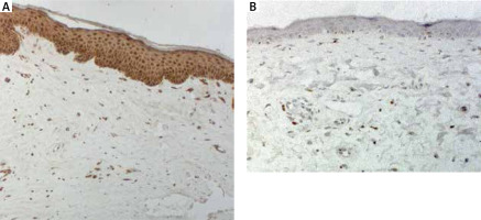
The quantitative evaluation of IL-17 expression in histological sections was presented as the mean number of positively stained cells in the field of view ± SD (Table 1). The number of cells showing IL-17 expression was significantly higher in VLS lesional skin as compared to normal skin of healthy controls (p < 0.0001). In VLS lesional skin, IL-17 was expressed in the epidermis and by cells within the inflammatory infiltrate in the upper dermis (Figure 1). The number of cells showing expression of IL-17 in the normal skin was relatively low. Representative immunohistochemical stainings of IL-17 in VLS lesional skin and in normal skin is presented in Figure 1.
Figure 1
Representative immunohistochemical stainings of IL-17 in VLS lesional skin (A), normal skin (B) (magnification, 200×)

Table 1
The quantitative evaluation of S100A7, IL-17 expression in lesional VLS skin, and normal skin of healthy controls (values expressed as the mean number of positively stained cells ± SD)
| Parameter | VLS (n = 21) | Normal skin (n = 10) | P-value |
|---|---|---|---|
| IL-17 mean ± SD | 88.97 ±20.08 | 30.14 ±14.17 | < 0.0001 |
| S100A7 mean ± SD | 67.04 ±16.16 | 14.49 ±18.31 | < 0.0001 |
Expression of S100A7
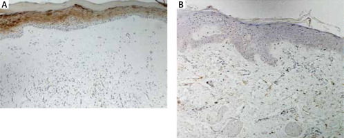
The quantitative evaluation of S100A7 expression in histological sections was presented as the mean number of positively stained cells in the field of view ± SD (Table 1). The number of cells showing S100A7 expression was significantly higher in VLS lesional skin as compared to normal skin of healthy controls (p < 0.0001) (Table 1). In VLS lesional skin, S100A7 was expressed by suprabasal keratinocytes in epidermis. S100A7 was also expressed by cells within the inflammatory infiltrate in the dermis (Figure 2). Representative immunohistochemical stainings of S100A7 in VLS lesional skin and in normal skin of healthy controls is presented in Figure 2.
Discussion
VLS/genital LS is a chronic progressive, lymphocyte-mediated inflammatory disease whose pathogenesis is complex and not fully elucidated. Various studies suggest a pivotal role of an autoimmune response and an immunological dysregulation in the development of LS. Both the primary and acquired immune response appear to be involved in the pathogenesis of this disease [1–3, 10]. The histological features of VLS comprise a dermal inflammatory infiltrate around the dilated capillaries composed mostly of lymphocytes (CD8+ T cells and to a lesser extent, CD4+ T cells) [2, 3, 12, 14]. In the inflammatory infiltrate in VLS, some other cells may be present e.g. mast cells distributed in the superficial dermis and macrophages present in the superficial and deeper part of the dermis (H1). The chronic inflammatory process is likely to induce the production of reactive oxygen species (ROS) which contribute to the skin damage and sclerosis [2, 32]. An overexpression of some proinflammatory cytokines such as TNF-α, IL-1α, IL-2 have been demonstrated in LS [2, 3, 10, 15]. In the present study we have investigated the expression of IL-17 in lesional skin obtained from female individuals with histologically confirmed genital LS (VLS). We found an increased expression of IL-17 in lesional skin of patients with VLS as compared to normal skin from healthy controls. IL-17 was overexpressed both in the epidermis and in the dermis, by cells within the inflammatory infiltrate. IL-17 is a potent proinflammatory cytokine that plays an important role in the development of the autoimmune inflammatory response. IL-17 is produced by multiple innate and adaptive immune cells including: Th-17 cells, NK cells, CD8+ T cells, innate lymphoid cells, γδ T-cells, and mast cells. Within the skin, IL-17 acts on cellular targets including keratinocytes, fibroblasts, neutrophils and endothelial cells and stimulate these cells for production of proinflammatory cytokines and chemokines e.g. TNF-α, IL-1, and IL-8 [16–18]. To the best of our knowledge, the expression of IL-17 has not been elucidated in the lesional skin of VLS. Our group [19] has indicated previously a significantly higher number of cells showing IL-17 expression in patients with cutaneous lichen planus compared to normal skin. The results of this study point toward the involvement of IL-17 in the pathogenesis of VLS. Overexpressed in lesional VLS skin, IL-17 may act on keratinocytes and activate these cells for increased production of proinflammatory mediators enhancing an inflammatory process in the skin. Moreover, IL-17 has been shown to act synergistically with other proinflammatory cytokines e.g. TNF-α to induce the expression of some antimicrobial proteins and to potentiate an inflammatory process [16, 17, 28].
S100A7 is an antimicrobial protein expressed by epidermal keratinocytes, in the inner root sheath of the pilosebaceous gland and in leukocytes: lymphocytes, neutrophils, and monocytes. S100A7 is encoded by the gene located in the epidermal differentiation complex (EDC) on chromosome 1q21 and is linked to the epidermal differentiation and inflammatory process. It regulates keratinocyte proliferation and differentiation and has proinflammatory activity, therefore may play a role in the pathogenesis of some chronic inflammatory skin diseases with epidermal abnormalities [24–26, 29, 31]. In the current study we have found an increased expression of S1007 in lesional VLS skin as compared to normal skin. S100A7 was overexpressed in keratinocytes in the suprabasal compartment of epidermis as well as in the dermal inflammatory infiltrate. To the best of our knowledge, the expression of S100A7 has not been much investigated in VLS/genital LS. Our results are consistent with one previously published study. Gambichler et al. [33] found an increased expression of S100A7 and human β-defensin (hBD)-2 in lesional LS skin, whereas the expression of other antimicrobial proteins: hBD-1, hBD-3, cathelicidin (LL-37) and RNase 7 was not significantly different than in normal skin. Some exogenous factors e.g. chronic irritation, trauma or alteration in skin microbiota are thought to trigger immune activation and inflammation in VLS. Koebner phenomenon was described in LS [2, 5, 33]. S100A7 has been identified as DAMP – ‘alarmin’. Upon mechanical and microbial stimuli, S100A7 may be released from keratinocytes and induce an inflammatory response. Moreover, IL-17 has been shown to act synergistically with TNF-α to induce expression of S100A7 in keratinocytes [25, 27–29]. Overexpressed in lesional VLS skin, S100A7 may promote epidermal abnormalities and potentiate an inflammatory process in the skin. S100A7 has been shown to stimulate keratinocytes for enhanced production of proinflammatory cytokines, such as TNF-α and IL-8 [28]. Further, S100A7 acts as a chemoattractant for leukocytes (e.g. lymphocytes, monocytes/macrophages and neutrophils) and its chemotactic activity is mediated by the multiligand receptor for advanced glycated end products (RAGE). S100A7 – RAGE interactions activate multiple intracellular signalling pathways, including nuclear factor κ-light-chain-enhancer of activated B cells (NF-κB) resulting in increased expressions of cellular adhesion molecules and proinflammatory cytokines [25, 27, 29, 31]. The results of our study may point toward the involvement of S100A7 in the pathogenesis of VLS.
Conclusions
VLS is a chronic, progressive debilitating disease whose pathogenesis is not completely known and has limited therapeutic options [1–3]. The results of our study may suggest the involvement of IL-17 and S100A7 in the pathogenesis of VLS. The better understanding of this disease may lead to the development of novel, effective therapeutic strategies.


