Introduction
Colon adenocarcinoma (COAD) is one of the most commonly diagnosed cancers of the gastrointestinal system. It is thought that more than 2.2 million new cases will be diagnosed and 1.1 million deaths will occur because of COAD by 2030. Unfortunately, despite adequate surgical therapies and chemotherapies, the 5-year survival rate of COAD patients is still poor. It is worth noting that the 5-year survival rate for patients diagnosed early is approximately 90%, whereas for patients with advanced diagnosis it is only 10%. This may indicate that metastasis is a critical cause of death for cancer patients [1–3]. Hence, there is an urgent need to identify novel prognostic biomarkers associated with metastasis, which can identify the high-risk patients and facilitate the timely application of appropriate individual treatments for COAD patients.
It has been widely reported that mitochondria are a known source of intracellular reactive oxygen species (ROS). Under physiological conditions they are constantly generated as by-products of aerobic metabolism in the mitochondria. During mitochondrial respiration, the tricarboxylic acid (TCA) cycle produces components with reducing properties, acting as a source for electrons. Electron transfer between mitochondrial electron transport complexes sets up a proton gradient for synthesis of ATP. It has been revealed that electrons can flee from the electron transport chain and react with oxygen molecules to form superoxide anions, which play a major role in a wide array of ROS [4]. It is worth mentioning that roughly 1–5% of the total oxygen consumed during the respiration process is transformed to superoxide radicals. Superoxide is highly reactive and toxic. For example, it may react with nitric oxide to form highly reactive peroxynitrite. These ROS are major factors associated with damage to mitochondrial lipids, proteins, and nucleic acids [5].
To counter the harmful effect of ROS, cells are possessed with an antioxidant defence system to detoxify ROS and avert them from their accumulation at high concentrations. The mitochondria-located manganese superoxide dismutase (MnSOD, SOD2) is known to convert superoxide to the less reactive hydrogen peroxide (H2O2), which may break down further into water and dioxygen by other enzymatic and non-enzymatic antioxidants. Because superoxide primarily emerges from mitochondria, mitochondrial MnSOD is thought to play a major role in detoxification of ROS [6,7].
Nevertheless, there is a little information about the expression of this protein in cancers of the digestive tract, especially in colon adenocarcinoma. It has been revealed that immunohistochemical expression of MnSOD was detected in colon adenoma and adenocarcinoma. However, there is no information about the influence of this protein on patients’ prognosis, especially those who did not receive any chemotherapies before surgical resection.
Aim
Therefore, the current study investigated the immunohistochemical expression of MnSOD in individuals living in Poland, who were diagnosed as colon adenocarcinoma patients, to assess its prognostic significance by correlating its expression with the clinicopathological factors and overall survival (OS).
Material and methods
Tissue samples
All colon adenocarcinoma samples were collected from patients who underwent radical surgery at the Municipal Hospital in Jaworzno. All patients were definitively diagnosed with colon adenocarcinoma and did not receive adjuvant chemotherapy or immunotherapy prior to surgery. The exclusion criteria were as follows: (1) history of previous malignant disease, (2) familial adenomatous polyposis, (3) inflammatory bowel disease, (4) preoperative anti-cancer treatment, and (5) evidence of distant metastasis. The clinicopathological characteristics obtained from the medical records were as follows: age, gender, location of tumour, grade of tumour differentiation, depth of invasion, regional lymph node involvement, operation record, treatment record, reoccurrence, and vital status at the last follow-up date. The follow-up period was 60 months (5 years).
The colon adenocarcinoma specimens belonged to 48 men and 49 women (mean age: 68; range: 33–89 years). Tumours were located in the proximal part of the colon in 50 (51.5%) cases and in the distal part of the colon in 47 (48.45%) cases. Two levels of differentiation were used to classify the grading as follows: well differentiated, 66 (68,04%) cases; and poorly differentiated, 31 (31.96%) cases (Table I).
Table I
Demographic, clinical, and tumour-related characteristics of patients included in the study (n = 98)
Immunohistochemical staining
For the immunohistochemical studies the paraffin-embedded specimens were cut into 4-µm-thick sections, fixed on Polysine slides, deparaffinised in xylene, and rehydrated through a graded series of alcohol. To retrieve the antigenicity, the tissue sections were treated twice with microwaves in a 10 mM citrate buffer (pH 6.0) for 8 min each. Subsequently, sections were incubated with rabbit monoclonal antibody to MnSOD (final dilution 1 : 400) (Abcam cat. number ab68155). For visualisation of protein expression, the sections were treated with the BrightVision detection system and Permanent AP Red Kit (Zytomed). Mayer’s haematoxylin was used to counterstain the nuclei.
The scores were assigned separately for the stained area and for the intensity of the immunohistochemical reaction. Quantification connected to the stained area of the tissue section was performed as follows: (1) < 33% of cells showed immunoreaction, (2) 33–66% of the cells had positive reaction to MnSOD, and (3) > 66% of the cells were positive. The intensity of the immunohistochemical reaction was quantified as follows: (1) absent or weak, (2) moderate, and (3) strong. Each tissue section was characterised by a final grade derived from the multiplication of the stained area and the intensity of the staining. The MnSOD expression was considered to be absent/low for grade 1; moderate for grades 2, 3, and 4; and strong for grades 6 and 9.
Statistical analysis
Statistical analyses were conducted using Statistica 9.1 (StatSoft, Poland). The clinical characteristics of the patients in relation to MnSOD immunoreactivity were assessed by performing the Kruskal-Wallis test and U Mann-Whitney test. The Kaplan-Meier method was used to study overall survival rate curves, and the log-rank test was used to compute differences between the curves.
Results
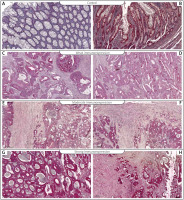
To investigate the prognostic utility of MnSOD expression, the immunohistochemical analysis was performed in colon adenocarcinoma tissues and adjacent non-pathological samples in patients diagnosed with colon adenocarcinoma. It should be pointed out that expression of MnSOD protein in non-pathological samples was described as weak or moderate. In samples of colon adenocarcinoma, the high, moderate, and weak levels of MnSOD protein were demonstrated as well (Figure 1). Among the colon adenocarcinoma samples, 24 (25%) showed weak immunohistochemical reaction, 27 (28%) demonstrated moderate immunoreactivity, and 46 (47%) showed strong expression. The relationships between the expression level and each clinicopathological parameter are summarised in Table II. As demonstrated, the level of the MnSOD immunohistochemical reactivity was not correlated with demographic factors including gender and age. The correlations were not also observed between MnSOD immunohistochemical reaction and clinicopathological factors (all p > 0.05; Table II).
Figure 1
Immunohistochemical expression of MnSOD protein in non-pathological colon samples (A, B) and in tumour tissue: C, D – weak expression of MnSOD protein in colon adenocarcinoma samples, E, F – moderate expression of MnSOD, G, H – strong expression of MnSOD

Table II
Correlations between MnSOD immunoexpression and clinicopathological characteristics in colon cancer patients
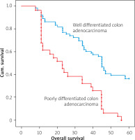
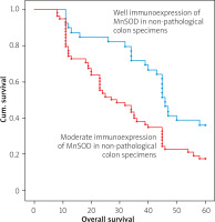
The Kaplan-Meier survival analysis showed that the overall survival rate in the group of patients with a low expression of MnSOD was not significantly longer than that for patients with a moderate or strong level of MnSOD immunoreactivity (p < 0.001). The low-MnSOD patients had an average survival time of 39.3 months (95% CI: 31.300–47.283), whereas the MnSOD moderate expression groups had an average survival time of 37 months (95% CI: 30.077–43.775). The strong-MnSOD patients had an average survival time of 35.5 months (95% CI: 33.134–40.536). The average survival time for all the patients was 36.8 months (95% CI: 33.134–40.536). Univariate log-rank test for overall survival in 97 patients with colon adenocarcinoma demonstrated that grade of tumour differentiation and MnSOD immunoexpression in non-pathological samples were significantly correlated with reduced OS (Table III). Moreover, a multivariate analysis demonstrated that grade of tumour differentiation (HR = 2.740; 95% CI: 1.864–4.027, p < 0.001) and MnSOD immunoexpression in non-pathological adjacent colon tissue (HR = 1.700; 95% CI: 1.073–2.694) were independent risk factors for worse survival (Table IV; Figures 2, 3).
Table III
Univariate log-rank test for overall survival in patients with colon adenocarcinoma
Table IV
Multivariate Cox regression analysis of overall survival in patients with colon adenocarcinoma
Discussion
MnSOD has been characterized as an antioxidant factor associated with the process of cancer development. Through the changing cellular processes, its up- and downregulation is clearly related to cancer development. A great number of studies have demonstrated that MnSOD expression was upregulated in many cancers. However, inhibition of MnSOD expression has been detected as well [8, 9]. The increased expression of MnSOD has been observed in cancer patients including lymphoma [10, 11], oral carcinoma [12–14], oesophageal squamous cell carcinoma [15], glioblastoma [16], ovarian carcinoma [17, 18], kidney cancer [19, 20], and colon cancer [21]. The results of our study demonstrated that expression of MnSOD was up-regulated in colon adenocarcinoma tissue in comparison to healthy ones. Interestingly, the level of MnSOD immunohistochemical expression was not correlated with clinicopathological factors and cancer progression. Also, the expression of MnSOD in colon adenocarcinoma patients with lymph node metastasis was not significantly higher than that observed in patients without metastasis. Moreover, the level of MnSOD expression in cancer samples was not correlated with the overall survival of patients. It should be pointed out that the level MnSOD expression in adjacent non-pathological tissue was correlated with reduced level of survival. Apart from the level of histological differentiation, the level of MnSOD in adjacent non-pathological mucosa was the second factor connected with poor prognosis. Our study is the first to demonstrate that expression of MnSOD is a factor that may be correlated with reduced survival of patients. But in this context our study has some limitations. Firstly, the patients are only from a European population living in Poland. Secondly, the small sample size has attenuated statistical power. Therefore, further studies are warranted to confirm our findings.
Connor et al. showed that the increased activity of Mn-SOD in bladder cancer causes a 3-fold increase in the invasiveness of this tumour. This indicates the enormous importance of this enzyme as a potential diagnostic marker and to monitor the patient’s condition and assess whether the tumour is entering an invasive phase. It seems therefore that elevated levels of MnSOD can be correlated with acquisition of invasive abilities during epithelial mesenchymal transition (EMT) [22]. Some studies revealed that MnSOD-dependent production of H2O2 increased the expression of matrix-degrading metalloproteinases (MMPs), forming permissive conditions for metastatic disease. Hempel et al. demonstrated that acquisition of metastatic phenotype was connected with increased expression of MnSOD in the 253J B-V cells [23]. It was also demonstrated in fibrosarcoma through MMP1 [24] in lung adenocarcinoma throughout the FoxM1–MMP2 axis [25]. A recent work has proposed a novel action of SOD2 as a sustaining factor in the Warburg effect. According to this study, SOD2 causes a metabolic shift to glycolysis in cancer cells by upregulating the AMP-activated kinase (AMPK) pathway, thus pointing out that H2O2 is also involved in cancer metabolic bioenergetics [26, 27].
Conclusions
The results of our study demonstrated that expression of MnSOD was up-regulated in colon adenocarcinoma tissue in comparison to samples without any pathological changes. The level of MnSOD immunohistochemical expression was not related to clinicopathological factors. It should be pointed out here that the level MnSOD expression in healthy tissues and the grade of histological differentiation was correlated with reduced OS.


