Introduction
In many professional articles using silver nanoparticles (AgNPs) is being discussed as an alternative to commonly used antimicrobial drugs [1, 2]. Several topical antibacterial products that are introduced to the market contain specifically designed AgNPs as a component for chronic wound treatment.
Both Gram-positive and Gram-negative bacteria as well as fungi can be isolated from chronic wounds. However, one of the most commonly isolated pathogens is Staphylococcus aureus. Due to antibiotic overuse these isolated strains are becoming multi-drug resistant, hence the search for an antimicrobial agent with a multifactorial effect but without a specific molecular goal has been carried out. AgNPs might be matching these criteria. Antibacterial activity of various AgNPs types has been proven on bacterial strains such as S. aureus, Streptococcus pyogenes, Pseudomonas aeruginosa and Escherichia coli [3]. Moreover, the antibiofilm, antifungal, anti-inflammatory and anti-neoplastic properties of AgNPs have been confirmed [4, 5].
Unfortunately, there are still many issues that need to be resolved and more research is needed before introducing AgNPs to large-scale therapeutics. Factors that could possibly affect the biological activity of AgNPs are still not fully understood due to lack of data. It is known that the smaller the nanoparticle is, the greater antimicrobial activity it exhibits. However, a smaller size is associated with increased cytotoxicity [6, 7]. Similarly, the nanoparticle shape is another feature that affects the antimicrobial activity [8] and it seems that coating factors may also play a role in NPs activity [9]. An important but notoriously overlooked issue is the stability of AgNPs solution. From all available synthesis methods only a few ensure 7-day kinetical stability of the product [10].
Aim
The aim of the study was to find stable AgNPs that could be effectively used in bacterial infection topical treatment with acceptable cytotoxicity levels. To achieve this several more detailed goals were set:
– Establishing kinetically stable AgNPs synthesis method and AgNPs characterization using physicochemical techniques,
– Evaluating AgNPs kinetical stability and maintaining AgNPs antimicrobial activity over time,
– Evaluating the coating factor influence on AgNPs antimicrobial activity,
– Evaluating AgNPs cytotoxicity against human keratinocytes (HaCat).
Material and methods
Synthesis and characterization of nanoparticles
Synthesis
For this purpose, a new method of AgNPs synthesis coated with long chain carboxylic acid (LCCA), esters of succinic acid and long chain alcohols (SAAE) was developed. Chemicals (except solvents) were purchased from Sigma Aldrich and were used without further purification. Solvents were purified with distillation.
The synthesis of silver nanoparticles using different chain length ammonium salt of carboxylic acid was based on modification of the literature procedure [11]. Carboxylic acids used during the synthesis have carbon chain length from two to sixteen carbon atoms. The monoester of the succinic acid with 1-decanol, 1-dodecanol, 1-tetradecanol, 1-hexadecanol were prepared according to the literature procedure [12]. The ammonium salts of carboxylic acids were prepared by mixing 1 mmol of acid dissolved in 10 ml of dioxane with 2 ml 1 molar ammonium hydroxide solution at room temperature, stirring at 40°C and evaporation to dryness. Solid ammonium salts were directly used for nanoparticles preparation without additional purification. Silver nanoparticles were obtained upon addition of 0.5 mmol carboxylic acid ammonium salts in 10 ml of water solution to stirred solution of 80 mg AgNO3 in 20 ml of water and mixing at room temperature. After 5 min to reaction mixture solution of 30 mg of sodium borohydride dissolved in 10 ml was added. The colour of the reaction mixture changed from light yellow to brown within 2 min. The above solutions were stirred for 1 h and used for subsequent experiments. For purification of nanoparticles, 10 ml of crude solution was taken into cellulose membrane (MEMBRA-CEL MC18 X 100 CLR) such that 70% by volume of the membrane was filled and kept in a 200 ml glass cylinder containing water (150 ml). The dialysis process was carried out for 48 h and water was replaced at least four times to remove all impurities and unreacted substrates [13].
AgNPs coated with LCCA were marked according to the number of carbon atoms in the particle, for example AgNPs coated with 12 carbon atoms carboxylic acids have the abbreviation C12. Those coated with esters of succinic acid and long chain alcohols were marked according to the number of carbon atoms in alcohol, for example: B10, B12, etc.
UV-visible spectroscopy
The synthesis of AgNPs was monitored by measurements of UV-vis absorption spectra (UV-VIS Hitachi U-1900, Hitachi High-Technologies Corporation) operated at a resolution of 0.5 nm. Analyses were taken immediately after adding the reducing agent and afterwards every 10 min up to the point where two identical spectres were observed. The same method was used to create NPs decay rate curves in a 14-day period. Furthermore, before every microbiological test, the spectrophotometer measurement was taken using Multiscan GO 3.2 to ensure NPs stability.
FTIR
Carboxylic acids and succinic acid monoesters both, before and after reduction were subjected to Fourier-transform infrared spectroscopy (FTIR; Jasco FP-6200, Japan) using the potassium bromide pellet technique and were exposed to an IR source of 500–4,000 cm–1.
DLS measurement
Freshly prepared AgNPs were subjected to particle-size analysis using dynamic light scattering.
Elementary analysis
Elementary analysis method was used to rate AgNPs elemental composition after the synthesis process. AgNPs solvents purified with dialysis were lyophilized (Lyovac GT2E 11, SRK Systemtechnik GmbH, Germany) and incinerated in elementary analyser (Vario EL III, Elementar Analysensysteme GmbH, Germany).
Microbiological assays
Reference strains of staphylococci, namely S. aureus ATCC 25923, S. aureus ATCC 12598, and S. aureus ATCC 6538 were acquired from the Polish Collection of Microorganisms. Clinical strains were isolated from the Department of Dermatology patients with chronic ulcerations in the course of chronic venous insufficiency. Strains were encoded to ensure patient anonymity (001W, 002W, 006W). Minimal inhibition concentrations (MICs) were determined by the broth microdilution method using 96-well polystyrene, in compliance with Clinical and Laboratory Standards Institute (CLSI) (2012) methods for dilution antimicrobial susceptibility tests for bacteria that grow aerobically; approved standard – Ninth Edition recommendations. For this purpose, bacteria at density of 5 × 10⁵ CFU/ml in Mueller-Hinton (MH) were exposed to a serial dilution of test AgNPs and incubated for 18 h at 37°C. The MIC was taken as the lowest concentration at which noticeable growth inhibition was observed. All test compounds were diluted in distilled water considering ion silver added during AgNPs synthesis, up to 1000 µg/ml concentration. All experiments were conducted in triplicate.
AgNPs cytotoxicity assays
HaCat keratinocytes were sown in 96-well plates (5 × 10³ cells/well) on DMEM HG broth (Sigma Aldrich) containing 10% FBS (Sigma Aldrich). The broth was swapped for serum-free after 24 h in CO2 incubator, and the cells were stimulated with AgNPs. After another 24 h at 37°C, the effect on proliferation was checked using XTT (2,3-bis(2-methoxy-4-nitro-5-sulfophenyl)-5-[(phenylamino) carbonyl]-2H-tetrazolium hydroxide) (Sigma Aldrich) test. Experiments were repeated 4 times.
Statistical analysis
Statistical analysis was performed using “R” program. The level of statistical significance was set at p-value < 0.05. Mann-Whitney U test was used to compare antimicrobial activity between various AgNPs and AgNO3, comparing anti-staphylococcal activity between various AgNPs themselves as well as cytotoxicity values. AgNPs stability over time was evaluated using Wilcoxon test.
Results
AgNPs synthesis
Multiple attempts were taken to synthesize AgNPs according to literature data. The resulting product AgNPs proved to be unstable and aggregated in several minutes or few hours at most. Very few synthesized AgNPs were stable enough to conduct microbiological tests [14, 15].
As a result, a new chemical synthesis method using sodium borohydride as a reductant factor and LCCA and SAAC as a coating factor was created. Systematically synthesized AgNPs were coated with carboxylic acids starting with citric acid (C2) to palmitic acid (C16). The stable products were achieved using LCCA (C11-C15) and for them a characteristic was performed. All synthetized AgNPs coated with SAAC were stable too.
AgNPs characteristics
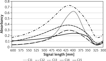
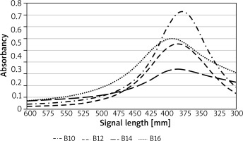
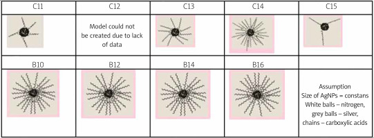
The UV-vis spectrum of all synthetized AgNPs exhibited bands characteristic for nanoparticles. UV-vis showed absorption shoulder at 370–420 nm for LCCA AgNPs and at 406–423 nm for SAAE AgNPs. UV-vis curves after the synthesis process are presented in Figures 1 and 2. DLS showed that AgNPs had an average size between 47 and 66 nm depending on the coating factor used. FTR results indicated that stabilizers used in the process did not oxidize and carboxylic bonds have been maintained but some structural changes have occurred. After coating AgNPs with lauric acid, some of the particles underwent dimerization. In case of C13, C14 and C15 an unsaturated bond was created in stabilizers’ structure. Similarly to C12, a formation of dimers was observed. It is worth mentioning that the stabilizer did not transform during AgNPs synthesis coated with SAAC (B10, B12, B14 and B16). Elementary analysis allowed estimating AgNPs coating density. For C11, the silver to carboxylic acid molar ratio was 11 : 1. On the surface of C15, very few carboxylic acid particles were present, coating density was small and unsaturated bonds were observed. The silver to carboxylic acid molar ratio was 7 : 1. The test on C12 provided unambiguous results. C13, C14 and B10, B12, B14, B16 analysis provided very different results. These NPs shown great surface stabilizer density since the carbon to silver atom ratio was close to 1 for C13 (1 : 3), C14 (1 : 22) and exactly 1 for B10, B12, B14, B16. Unsaturated bonds were not present for this AgNPs group. Elementary analysis also detected nitrogen atoms in some of the AgNPs structure, most noticeably in those with higher density: C14, B10, B12, B16. The presence of nitrogen in the structure clearly indicates that the ammonium ion is incorporated into the surface of AgNPs. Stabilization effect of amines on nanoparticles stability was already mentioned/discussed in the literature. AgNP models presented in Figure 3 were based on obtained results.
AgNP kinetical stability assessment
Spectrophotometer readings were taken 14 days after the synthesis process was complete. AgNPs coated with short chain acids starting with acetic acid (C2) up to pelargonic acid (C9) aggregated during the first 24 h after synthesis. AgNPs coated with capric acid (C10) shown kinetical stability for 4 days. AgNPs coated with long chain carboxylic acid (LCCA AgNPs): undecyclic (C11), lauric (C12), tridecyclic (C13), myristic (C14) and pentadecyclic (C15) as well as all SAAE (B10-B16) were stable throughout the 14-day experiment.
SAAE AgNPs and LCAA C11-C15 AgNPs could be used in biological research since they were stable for at least 3 days (minimum biological research timeframe). Table 1 represents solvent stability during 14-day UV-vis spectroscopy readings after AgNPs synthesis.
Table 1
The stability of AgNPs water solutions based on spectrophotometer measurements
Gradual AgNPs aggregation could impact biological effectiveness. Whether AgNPs kinetical stability corelates directly to maintaining their antimicrobial characteristics through time is unknown. To verify this hypothesis microbiological research on synthesized solvents with MIC indicators using serial dilution methods in time intervals was conducted. Average MIC marks were statistically compared for all S. aureus strains. MIC values decline for LAAC AgNPs was not statistically significant after 14 days after synthesis. After 4 weeks MIC values for C12 and C15 showed a statistically significant increase what indicates loss of AgNPs antimicrobial characteristics in time. Statistical analysis results are shown in Table 2.
Table 2
Changes in anti-staphylococcal activity of LCCA coated AgNPs in time with statistical analysis
AgNPs SAAE high activity even after a few months after synthesis was discovered during preliminary research. Solvent activity was compared after 3 and 6 months. With a 95% statistical significance level it was proven that SAAE AgNPs maintained anti-staphylococcal activity for 12 weeks. After 24 weeks, only B16 solvent exhibited a statistically insignificant MIC value decline comparing to values taken immediately after synthesis. Statistical analysis results are shown in Table 3.
Table 3
Changes in anti-staphylococcal activity of SAAC coated AgNPs in time with statistical analysis
Anti-staphylococcal activity of AgNPs
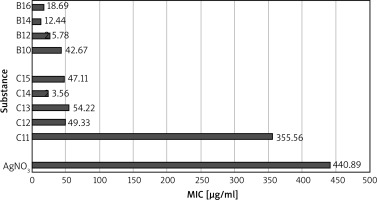
The antimicrobial activity of AgNPs was tested on six S. aureus strains. At the beginning of the test, MIC values were obtained for AgNO3 since it has been used in treatment for years, with the average MIC value of 440.89 µg/ml (test was conducted on all six strains). For the first time, systematic testing was conducted to assess how increasing the stabilizer carboxylic chain length affects AgNPs antimicrobial properties. Figure 4 shows comparison between AgNPs antimicrobial properties and silver nitrate as a reference. MIC values for C11 and AgNO3 did not have a statistically significant difference in antimicrobial activity (p = 0.11). However it was determined that AgNPs coated with carboxylic chains with > 11 carbon atoms and SAAE show significantly better antimicrobial properties than AgNO3. Moreover, it was noted that antimicrobial activity corelates with carbon chain length what can be used to modify AgNPs biological activity.
AgNPs cytotoxicity evaluation
Conducted research clearly shows that SAAE AgNPs have the highest kinetical and microbiological stability. They also have the strongest bacteriostatic effect on S. aureus strains hence they are the most likely AgNPs to be used topically as germicidal substances.
Therefore, cytotoxicity tests were performed only for SAAC AgNPs. B16 AgNPs had the highest cytotoxicity levels, 50% of cells have died in a 6.3 µg/ml concentrated AgNPs solution. This concentration is almost two times lower than the average MIC values for S. aureus at 16.89 µg/ml. Concentration cytotoxic for keratinocytes in case of B10 (IC50 = 50 µg/ml vs. MIC = 42.67 µg/ml), B12 (IC50 = 50 µg/ml vs. MIC = 25.78 µg/ml) and B14 (IC50 = 39.9 µg/ml vs. MIC = 12.44 µg/ml) turned out to be greater than solution’s average MIC values, what makes only these 3 AgNPs viable for further AgNPs treatment research wherein B10 has a very narrow therapeutical index between bacteriostatic and cytotoxic concentration.
Discussion
Considering increasing bacterial resistance to antibiotics, AgNPs antibacterial properties are being intensively researched for industrial and medical use. There is a number of review articles about AgNPs chronic wound treatment indicating AgNPs as a perfect alternative to currently used products dedicated to the treatment of ulcers [2, 16–22]. Whether these new products are stable enough to be used in local antimicrobial applications is still up for discussion.
Kalashnikova et al. indicate that NPs solvent stability is a key factor when considering AgNPs practical use [2]. Patakfalvi and Dékány proved that a citric acid stabilizer (coating factor) prevents AgNPs from aggregating [23]. This effect was explained by Labille and Brant who indicated that nanoparticles naturally seek a more thermodynamically favourable state and aggregate [22]. Paredes et al. pointed out that cation surfactants are highly effective as AgNPs solvent stabilizers [24]. Size increase causes the particles to lose their characteristics, typical for “nano” structures [22]. Li et al. thesis contains research on the stability of PVP, Tween (polymer) and citric acid coated NPs. These NPs were deemed unstable over the course of the experiment (960 s) [10, 25]. It is worth mentioning that conducting basic biological research requires at least 3 days. Authors acknowledged coating factors’ effect on AgNPs stability and found that oleic acid coated AgNPs stood out in terms of stability and maintained their characteristics for a few months [15]. Oleic acid’s complex structure should be taken into consideration as it contains a double bond. Due to this fact, it is difficult to identify the exact feature that provides the stabilizing effect: carbon chain length, double bond presence or its position in the chain etc. Considering multiple factors that can affect AgNPs behaviour it was deemed necessary to perform systematic research on carbon chains length influence on AgNPs. A new AgNPs coated with simple unsaturated carboxylic acids synthesis method was developed. It was proven that extending the carbon chains length by adding one carbon atom affects AgNPs stability as well as the final product’s physical state. AgNPs stable for at least 14 days were coated with acids: undecanoic (C11), lauric (C12), tridecanoic (C13), myristic (C14) and pentadecanoic (C15) as well as all SAAE. The stabilizer chemical structure characterized by long carbon chain has a potential for further molecule modification for example by adding antibiotics. This opens up new perspectives for further synthesis research of highly specific acting AgNPs.
Another important aspect when considering AgNPs antimicrobial usage besides their solvent stability is maintaining their antimicrobial properties in time. There is no research on this subject in available literature. Most LCCA AgNPs maintain anti-staphylococcal properties for 4 weeks. SAAE AgNPs maintain anti-staphylococcal properties for 3 months and in case of B16 even for 6 months. Finally, coating factors’ impact on biological activity needed to be verified. Similarly synthesized, with a similar shape and size AgNPs were chosen for the test. Microbiological tests showed that anti-staphylococcal effect depends on the number of carbon atoms in the carboxylic chain. AgNPs MIC value analysis shows that the more carbon atoms are present in the carboxylic chain the more potent the substance is: C11 < C12 < C13 < C14 with the exception of C14 > C15. AgNPsB can be also described this way as: B10 > B12 > B14 wherein statistically significant loss of effectiveness was not observed between B14 and B16. Moreover, between C14 and SAAE AgNPs group there was no statistically significant difference in antistaphylococcal activity. Conducted physicochemical analysis allowed creating AgNPs models. The analysis showed how carbon chain extension affects density of coating of the AgNPs structure. Based on physicochemical analysis results and microbiological research a hypothesis can be made that AgNPs’ coating degree with stabilizer affects its biological activity. He et al. explain the interaction between NPs and biological films indicating adsorption as one of the ways NPs enter cells. Authors pointed out fat solubility of substances is key in adsorption process. The more densely coated and more lipophilic NPs structure the easier it is to penetrate the lipid bilayer [26]. However further research is needed to prove this hypothesis.
Conclusions
Kinetically stable AgNPs synthesis method was established for AgNPs coated with long chain carboxylic acid and AgNPs coated with esters of succinic acid and long chain alcohols. Assay results indicated the coating factors effect on kinetical stability and biological activity of AgNPs. From all examined AgNPs types only two substances were sufficiently kinetically stable and had high bacteriostatic effectiveness against S. aureus and at the same time low enough cytotoxicity levels for epidermis cells so that they can be considered for topical antiseptic usage: B12 and B14 AgNPs.


