Bullous pemphigoid (BP) is an autoimmune disease characterized by the production of antibodies reactive with the basement membrane band antigens BP180 and BP230. The interaction of antibodies with these antigen results in the formation of sub-epidermal blisters. The pathogenesis of BP is unclear, although specific triggers for BP have been identified such as drugs, scalds, electrical injury, surgery, and ultraviolet irradiation [1]. Herein, we report a case of BP that occurred at the site of a closed fracture.
A 53-year-old female presented with erythema and blisters on the skin of her left wrist. A closed bone fracture of the left wrist had occurred 1 month prior to presentation at our hospital and the 1-month period coincided with the erythema and blisters. Treatment for that period was 0.05% halometasone cream, which was marginally effective and she used only a small splint immobilization. She had a 5-year history of hypertension that was treated with nifedipine sustained-release tablets, administered orally, 20 mg daily. She had no other comorbidities and was not using any other drugs.
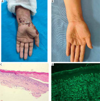
Physical examination showed scattered dark erythema, blisters, and crusts ranging from 4 mm to 1 cm in diameter on the left wrist and back of the hand. The wall of blisters was tense and the Nikolsky’s sign was negative (Figure 1 A). Laboratory investigation found a high eosinophil count (0.77 × 10⁹/l, reference range 0.02–0.52 × 10⁹/l) and eosinophil proportion (15.5%, reference range 0.4–8%), high levels of anti-BP180 autoantibodies (BP180-Ab) 67.4 U/ml and anti-BP230 autoantibodies (BP230-Ab) > 200 U/ml by ELISA diagnostic method (EUROIMMUN). Antibodies reactive with desmoglein (DSG)1/3 and type VII collagen were not present. Liver and kidney function, blood glucose, blood lipids, electrolytes, immunoglobulin, complement, and C-reactive protein levels were all normal.
Figure 1
A – Before treatment: scattered dark erythema and blisters ranging from 4 mm to 1 cm in diameter on the left wrist and the back of the hand. B – After treatment: the lesions on the left wrist and back of the hand subsided. C, D – Histopathology showed sub-epidermal vesicles with abundant eosinophil infiltration in the papillary dermis (C: hematoxylin and eosin staining (HE) 40×). Direct immunofluorescence (DIF) showed a clear band of immunoglobulin G (IgG) and C3 deposition at the dermo-epidermal junction (D: HE 100×)

Skin biopsy of the left hand was performed and histopathology showed a sub-epidermal vesicles with abundant eosinophil infiltration in the papillary dermis. Direct immunofluorescence (DIF) showed a clear band of immunoglobulin G (IgG) and C3 deposition at the dermo-epidermal junction (Figures 1 C, D). Salt-split indirect immunofluorescence (IIF) found IgG autoantibodies reacting along the epidermal side of the split skin. It was human skin, and we used our own reagents. Based on this evidence, a diagnosis of BP was made. The patient was started on oral minocycline, 100 mg twice a day, and nicotinamide tablets, 500 mg three times a day. Her lesions completely resolved during a 2-month treatment period (Figure 1 B), when treatment was stopped. BP180-Ab and BP230-Ab were not found at her 8-month follow-up. There was no further symptom recurrence reported at her 10-month follow-up.
As noted above, occurrence or aggravation of BP has been related to specific triggers such as drugs, physical factors, vaccines, infection, and transplantation [1]. Among the physical factors, trauma, surgical incision, and ultraviolet irradiation are relatively common. Mai et al. [2] reported that BP induced by physical factors was more common in younger individuals, with the incidence greater in women. Skin lesions are typically limited to the injured site.
In this case, BP occurred at a closed fracture site, in which the skin surface was intact without damage. A previous report [3] described a 61-year-old woman with a right femur closed fracture who developed BP after femur fracture surgery. Lesions occurred at the site of the surgical incision, thus BP was thought to be caused by surgical trauma. In this case, we suspected that the BP trigger was not a skin element but rather the closed fracture itself, which differs from the previous report.
The pathogenesis of BP, induced by trauma, is unclear. Tissue destruction caused by trauma may lead to basement membrane band antigen exposure. Such antigen exposure may induce an inflammatory response and the production of circulating antibodies, which damage tissues. The damaged tissues may release a variety of pro-inflammatory factors, recruiting inflammatory cells and circulating antibodies that activate granulocytes and the complement system, resulting in the formation of blisters [4]. Alternatively, the epitope spreading phenomenon may occur, which has been demonstrated in both experimental murine BP and human BP [5]. Herein, we report an interesting phenomenon, the specific pathogenesis of which requires further investigation. We plan to collect more patients with BP triggered by a closed fracture and to compare the immunological findings between closed fracture-induced BP and classic BP.


