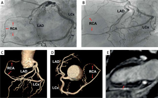
A 62-year-old woman, with a history of smoking, presented to our cardiology service with acute chest pain. Her electrocardiogram (ECG) recorded ST-segment depression in leads V3 → V6, and blood tests demonstrated elevated high-sensitive troponin-I levels (maximum of 2.579 pg/ml, normal < 15.6 pg/ml), indicating a non-ST-elevation myocardial infarction (MI). Transthoracic echocardiography showed no wall motion abnormalities. Coronary angiography was performed, revealing a non-obstructive single coronary artery originating from the left sinus of Valsalva. In particular, the right coronary artery (RCA) arose from the distal part of the left circumflex artery (LCx), continuing into the right atrioventricular sulcus (Figures 1 A, B, arrows). Coronary computed tomography angiography revealed an anterior course of the RCA to the great vessels (type L- IA) [1], precluding any chance of compression (Figures 1 C, D, arrows).
Figure 1
Coronary angiography showing a non-obstructive single coronary artery originating from the left sinus of Valsalva. The right coronary artery (RCA) arises from the distal part of the left circumflex (LCx) continuing into the right atrioventricular sulcus (A, B, arrows). Coronary computed tomography angiography deciphered an anterior course to the great vessels (C, D, arrows). Cardiac magnetic resonance imaging depicts a small sub-endocardial area of late gadolinium enhancement in the mid-inferior and inferolateral wall (E, arrow)

To differentiate between MI with non-obstructive coronary arteries (MINOCA) and myocarditis, cardiac magnetic resonance imaging was performed. A small subendocardial area of late gadolinium enhancement was observed in the mid-inferior and inferolateral wall (Figure 1 E, arrow), establishing the diagnosis of MINOCA. She was treated with metoprolol, aspirin and atorvastatin followed by ECG normalization and complete symptom resolution.
Single coronary artery is a rare congenital anomaly, especially the L-IA endotype, where only one coronary artery arises from a single coronary ostium, supplying the entire heart [1, 2]. Apart from the endotypes which predispose to compression, no definite connection to myocardial ischemia has been proposed [1]. MINOCA accounts for 6% of all MIs, affecting mostly younger females without traditional risk factors [3]. To the best of our knowledge, this is the first description of MINOCA in a patient with a single coronary artery.


