Introduction
Coronary artery disease (CAD) is the foremost single cause of mortality and loss of disability-adjusted life years (DALYs) globally and a large percentage of this burden is found in low and middle income countries. It accounts for nearly 7 million deaths and 129 million DALYs annually and is a huge global economic burden. [1].
CAD is characterized by the presence of atherosclerosis in coronary arteries. The underlying pathophysiologic mechanisms for these syndromes begin with the process of atherosclerosis, which develops and progresses for decades prior to the acute event. Management of CAD has 2 main goals: to reduce symptoms due to ischemia and to prevent myocardial infarction and death. Medical management is pivotal in all patients with CAD and aims to identify and treat any associated diseases that can precipitate angina by increasing the myocardial oxygen demand (such as tachycardia and hypertension) or by decreasing the amount of oxygen delivered to the myocardium (such as heart failure, pulmonary disease, or anemia) and to manage CAD risk factors as well as to prevent myocardial infarction (MI) with lifestyle changes and pharmacological treatment.
The methods of revascularization are well established for CAD: coronary artery bypass grafting, introduced in 1968, and percutaneous coronary intervention.
The benefits of coronary revascularization in reducing cardiac events and death have been widely accepted in the context of acute coronary syndromes with ST-segment elevation MI and non-ST-segment elevation MI.
Since the aim of the surgical procedure is not only restoration of blood flow in coronary arteries but also improving function of myocardium, identification of dysfunctional but viable myocardium suggesting potential reversibility of myocardial function is of clinical importance. Unfortunately, restitution of coronary blood flow does not always mean improvement in myocardial function. Imaging techniques such as myocardial uptake of F-18 fluorodeoxyglucose with positron emission tomography (PET) or thallium-201 reinjection scintigraphy has been proved clinically useful for predicting which myocardial segments will demonstrate improvement after revascularization. Tc-99m-MIBI SPECT with very good physical characteristics and passive diffusion driven by mitochondrial transmembrane potential closely reflects blood flow distribution [2].
Aim
In our study we aimed to evaluate patients with coronary artery disease who were planned for coronary artery bypass grafting (CABG), using 2D echocardiography and myocardial perfusion imaging (MPI) both preoperatively and postoperatively in an attempt to assess the myocardium that might have undergone reversible insult from irreversible insult. In doing so we also wanted to evaluate the utilisation of MPI as a routine investigation for patients with lower ejection fractions that might benefit from revascularisation by identifying viable myocardium. During the course of our study we also aimed to study the demographics of the patients undergoing coronary artery bypass grafting, the vessel character, whether diffusely diseased or focally diseased, associated comorbidities and comparison of the ejection fraction using 2D echocardiography, MPI and the improvement in both of these, if any, postoperatively.
Material and methods
This prospective observational study was conducted at our tertiary care center. Institutional ethical committee approval was obtained before the start of the study (UNMICRC/CVTS/2019/16). All the patients having coronary artery disease planned for CABG between December 2019 and November 2021 were included. Demographic details of all the patients were collected. All these patients underwent routine coronary angiography along with myocardial perfusion imaging preoperatively. Their vessel character was noted, based on coronary artery angiography preoperatively and broadly classified into diffuse vessel disease or focal disease. Postoperatively the patient was evaluated using 2D ECHO and MPI. MPI was done 3 months after the operative procedure. All grafting and intraoperative assessment was done by the same senior cardiac surgeon.
All the patients who had coronary artery disease planned for coronary artery bypass grafting (CABG) between December 2019 and November 2021 were included in the study.
The patients requiring emergency CABG and concomitant valvular and vascular surgery were excluded from the study.
Preoperative management:
All routine investigations,
Coronary angiography,
ECG,
2D Echo,
Myocardial perfusion imaging,
MPI Protocol.
Postoperatively in follow up after a year:
2D ECHO,
MPI.
MPI:
Segmental analysis,
Ejection fraction (EF),
Perfusion deficit (PD),
Summed rest scores (SRS).
Segmental analysis by 99mTc-Sestamibi: the American Heart Association, American College of Cardiology, and Society of Nuclear Medicine have defined the standards for plane selection and display orientation for serial myocardial slices generated by SPECT imaging with respective nomenclature, i.e. short, vertical long, and horizontal long axes [3].
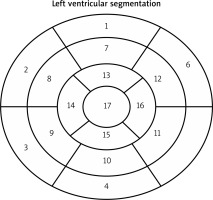
The 17 segment approach: For regional analysis of the left ventricular function or myocardial perfusion, the left ventricle is divided into equal thirds perpendicular to the long axis of the heart. This generates 3 circular basal, mid-cavity, and apical short-axis slices of the left ventricle (Figure 1).
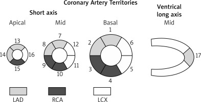
Although there is a tremendous variability in the coronary artery blood supply to myocardial segments, it was believed to be appropriate to assign individual segments to specific coronary artery territories. The assignment of the 17 segments to one of the 3 major coronary arteries is shown below. The greatest variability in myocardial blood supply occurs at the apical cap, segment 17, which can be supplied by any of the 3 arteries. Segments 1, 2, 7, 8, 13, 14, and 17 are assigned to the left anterior descending coronary artery distribution. Segments 3, 4, 9, 10, and 15 are assigned to the right coronary artery when it is dominant. Segments 5, 6, 11, 12, and 16 are generally assigned to the left circumflex artery (Figure 2).
Assessment of myocardial salvage can be carried out more accurately by SPECT or CMR, which measures valuable parameters such as, area at risk (AAR) and final infarct size (FIS), from which myocardial salvage and myocardial salvage index can be derived.
Scoring left ventricular myocardial perfusion during rest using 17 segments: For the semi-quantitative evaluation of left ventricular perfusion, summed rest score (SRS) is used to calculate the perfusion score considering both the extent and severity of ischemia in relation to the 17 segments of the polar map. Normal perfusion (as compared with average gender-specific data from the population of healthy individuals) is indicated on the scale as a score of 0 (normal perfusion in relation to the control group). Mild and moderate perfusion impairment is indicated by 1 and 2 points, respectively. A score of 3 points indicates significant perfusion impairment, while a score of 4 points is used to indicate total impairment, meaning practically no perfusion. It is estimated that a left ventricular perfusion deficit with a score of 1 or 2 indicates that isotopic activity in this segment is approximately 60% in comparison to the area of radiotracer accumulation specified as 100%. Three points indicate radiotracer activity between 40% and 60%, while 4 points indicate radiotracer activity below 40% in relation to the area with 100% activity. Scoring perfusion disturbances in resting examinations is also useful for evaluating myocardial vitality.
Statistical analysis
Statistical analysis was carried out using SPSS version 20.0 software (SPSS Inc, USA). These data were presented as mean ± SD or proportion as appropriate. Number and percentage was used for categorical data and for continuous variables. Independent student ‘t’ tests and paired sample T test were used. The p-value less than 0.05 was considered to be significant.
Results
This study was conducted over a period of two years. We evaluated 50 patients who underwent CABG, their evaluation was done through 2D ECHO and myocardial perfusion imaging preoperatively and they were followed up after a period of 1 year and evaluated again using 2D ECHO and MPI. One patient died during the perioperative period, due to uncontrolled hypertension leading to dissection requiring surgery and succumbed to MODS and Sepsis and for the same reason was excluded from the study.
Out of 49 patients, 38 were male and 11 were female patients. The mean age of the patients participating in the study was 57.7 years. 34 of the patients participating in the study were diabetics and 17 were hypertensives, and 17 patients had both hypertension and diabetes mellitus.
Patients underwent coronary angiography preoperatively and out of the patients evaluated the majority had triple vessel disease (39) and 9 had double vessel disease and 1 patient had single vessel disease (Table I).
Table I
Demographic details
| Variables | N (%) |
|---|---|
| Mean age | 57.7 |
| Sex: | |
| Male | 39 (78%) |
| Female | 11 (22%) |
| Diabetes mellitus | 35 (70%) |
| Hypertension | 17 (34%) |
| Diffuse disease | 43 (86%) |
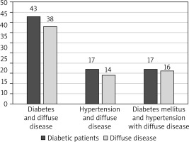
Amongst the patients who participated in the study it was found that most of the patients who had comorbidities (DM and hypertension) had associated diffusely diseased vessels. Out of the 43 diabetic patients, 5 patients had focal lesions and out of the 17 hypertensive patients, 3 patients had focal lesions. Only one patient with both diabetes and hypertension had focal disease (Figure 3).
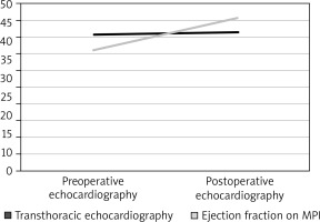
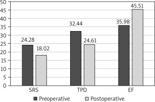
Patients were evaluated using transthoracic 2D echo preoperatively and postoperatively. Mean ejection fraction preoperatively was 40.6 ±9.72% and postoperatively it improved to 41.32 ±10.64%. Similarly, patients’ ejection fraction was calculated using MPI and an average improvement from 35.98 ±12.72% to 45.51 ±12.61% (p < 0.0001) (Table II, Figure 4).
Table II
Preoperative and postoperative EF difference
The data were analysed before and after CABG during MPI scanning and SRS and perfusion deficit was calculated.
SRS was calculated and an improvement was noted from 24.28 ±8.47 to 18.02 ±8.75 (p < 0.0001). Total perfusion deficit was calculated and was found to have reduced from 32.44 ±11.98 to 25.61 ±12.23 (p < 0.0001) (Table III, Figure 5).
Table III
Preoperative to postoperative summed rest score and perfusion defict differences
When comparing the changes in the Summed Rest Scores and Total Perfusion Deficit in patients who had diffuse disease, we found that SRS increased from 24.83 ± 8.75 to 18.73 ± 9.0 and the TPD reduced from 32.92 ± 11.82 to 25.07 ± 11.96. The change was greater in patients with focal lesions with a change in SRS from 22 ± 7.48 to 15 ± 7.39 and TPD from 31.5 ± 18.57 to 26 ± 18.61. The p value was however not significant.
Discussion
In our study conducted over a course of 2 years, patients undergoing CABG were evaluated preoperatively and postoperatively using myocardial perfusion imaging as a modality of investigation. Our aim was to attain a demographic profile of the patients being operated and their comorbidities. Preoperative and postoperative 2D echocardiography and preoperative and postoperative MPI. Fifty patients consented to participate in the study. One patient expired in the immediate postoperative period and was excluded from the study.
Out of the 49 patients who were followed up we found that 38 males and 11 females participated in the study. Indians are said to have the highest CAD rates. [4]. Cardiovascular disease does develop in women but is said to develop 7–10 years later than in men, however it is still a major cause of death in females over 65 years [5]. The mean age of the participants in the study was 57.7 years. In their study ‘Gender- and age-related differences in clinical presentation and management of patients with stable coronary artery disease’ by Ferrari et al. on evaluating 33280 patients, the mean age was 64 years and 22% of the patients were females [6].
Majority of the patients were diabetics (69%) and 34% of the patients had hypertension. 34% of the patients had both hypertension and diabetes. Diabetes has become a challenge for India with a prevalence of 8.8% in the 20–70 years age group. The prevalence of CAD in Indians living in India is 21.4%. Hypertension is attributable to 10.8% of all deaths in India [7]. Its prevalence has been on a steep rise over the past three decades both in urban and rural areas. This burden is expected to rise twice from 118 million in 2000 to 213.5 million by 2025. It was estimated that 16% of CAD, 21% of PVD, 24% of AMI and 29% of strokes are attributable to hypertension. Indians consume high carbohydrate diets with uneven dietary patterns.
In our study the patients were also evaluated using 2D echocardiography preoperatively and a year after the procedure and we found that there was an increase in the ejection fraction from 40.6 ±9.72% to 41.32 ±10.64%,which was statistically significant (p < 0.05). In another same study we have found that improvement of ejection fraction after coronary artery bypass grafting surgery in patients with impaired left ventricular function was evaluated in 40 patients of the average age being 59.8 years out of which 81.3% were male and 18.8% female [8]. They found one-vessel disease present in 2.5% (1/40) of patients, two-vessel disease in 40% (16/40), three-vessel disease in 42.5% (17/40) and four-vessel disease in 15% (6/40) of patients. Left ventricular ejection fraction assessed preoperatively was 18–27% and postoperatively it was improved to 31.08% in a period of 30 days. They concluded that in patients with left ventricular dysfunction, coronary artery bypass grafting can be performed safely with improvement in quality of life and in the left ventricular ejection fraction. Similar results were seen in the study ‘Left ventricular function outcome after coronary artery bypass grafting, King Abdullah Medical City (KAMC) – single-center experience’ by Khaled et al. [9] who examined 110 patients with left ventricular ejection fraction (LVEF) < 50% who underwent CABG with a mean age of 56.1 ±12.2 years old. They classified the patients into two groups: group I, 76 (69%) patients with LVEF > 35%, and group II, 34 (31%) patients with LVEF < 35%. There was a significant improvement in LVEF post-surgery (p = 0.05) in both groups with no significant difference recorded for in-hospital mortality rate among patients in different groups. They concluded that there is a significant improvement of LV systolic function after CABG and hence better survival rate. DM, significant diastolic dysfunction, and perioperative insertion of IABP are predictors of mortality after cardiac surgery and special care should be provided to such patients to improve their outcome.
During MPI, ejection fraction was calculated both preoperatively and postoperatively and an improvement was seen in the ejection fraction from 35.98 ±12.72% to 45.51 ±12.61% (p < 0.05). An increase in LVEF after coronary revascularization could be due to hibernation, i.e. resting ischaemia in viable myocardium. Stunning has also been suggested as an explanation. We observed improved LVEF values in our patients.
In our study during MPI we calculated the SRS and the perfusion deficit in our patients and we found a statistical difference in both parameters postoperatively. SRS improved from 24.48 ±8.47 to 18 ±8.75 and total perfusion deficit was seen to improve from 32.44 ±11.98 to 24.61 ±12.23.
Areas that were non-viable did not improve but areas that were partially and completely reversible improved postoperatively after coronary artery bypass grafting. On calculating the perfusion index (perfusion deficit divided by the number of areas assessed) we noticed an improvement in them too. Similar results were obtained by Paluszkiewicz et al. who evaluated 32 patients using dobutamine stress echocardiography and Tc-99m-MIBI SPECT and reduced significantly in their study too (2.19 ±0.71 vs. 1.93 ±0.70; p = 0.0008) [4].
In their study ‘Beneficial effect of coronary artery bypass grafting as assessed by quantitative gated single-photon emission computed tomography’, Hida et al. used gated SPECT to assess the benefit of CABG in patients with coronary artery disease; 47 of those patients were evaluated before and 5 months after CABG [10]. As a result of coronary revascularization, a significant improvement was observed in the global ejection fraction (50 ±12 → 53 ±11%; p < 0.05), average regional perfusion scores at rest (0.6 ±0.6 → 0.3 ±0.4; p < 0.0001), average regional wall motion score (0.9 ±0.7 → 0.7 ±0.5; p < 0.05), and end-diastolic wall thickness (8.1 ±1.3 → 8.6 ±1.5 mm; p < 0.0005) all improved significantly. Even in 34 non-revascularized territories, the average regional reversible defect score (0.5 ±0.7 → 0.2 ±0.5; p < 0.03), average regional wall motion score (0.8 ±1.1 → 0.5 ±1.0; p < 0.03). These results indicate that improvement in myocardial ischemia, hibernation and left ventricular function with CABG can be assessed in detail with gated SPECT [11]. Similar results were obtained in our study, when comparing the ejection fraction and the perfusion scores using SRS.
In their study ‘Myocardial perfusion correlates with improvement of systolic function of the left ventricle after CABG. Dobutamine echocardiography and Tc-99m-MIBI SPECT study’, Paluszkiewicz et al. [4] aimed to assess the effect of surgical revascularization (coronary artery bypass grafting (CABG)) on systolic function and perfusion of the left ventricle using dobutamine echocardiography (DE) and Tc-99m-MIBI SPECT (SPECT). There were 32 patients with a mean age of 52.2 ±7.2 years in whom DE and SPECT were performed before and 3–4 months after CABG using standard protocols. Wall motion score index (WMSI) and perfusion index (PI) were calculated. Perfusion index was defined as the net SRS divided by the number of regions assessed. They noticed a significant improvement of WMSI at rest (1.44 ±0.46 vs. 1.33 ±0.41; p = 0.03) as well as after the maximal dose of Dobutamine (1.49 ±0.42 vs 1.39 ±0.44; p = 0.02) was observed after CABG as compared to preoperative examination. A similar relation was observed during SPECT study. Perfusion index diminished significantly after revascularization during rest acquisition and after Dipyridamole administration (2.73 ±0.73 vs. 2.20 ±0.69; p = 0.0001) as compared to preoperative examination. We found a correlation between PI and WMSI at rest before CABG (R = 0.46; p = 0.01), PI after Dipyridamole and WMSI after the maximal dose of Dobutamine before CABG (R = 0.37; p = 0.04), PI and WMSI at rest after CABG (R = 0.39; p = 0.03), PI after Dipyridamole and WMSI after Dobutamine after CABG (R = 0.38; p = 0.03). Even though this study included stress SPECT as a component of the study and our study was at rest we found a similar improvement in our patients when comparing the SRS preoperatively and postoperatively and the perfusion deficit increased significantly post-surgery.
In their study ‘Electrocardiographic gated 99mTc-MIBI SPECT for functional assessment of patients after coronary artery bypass surgery: comparison of wall thickening and wall motion analysis’, Junichi Taki et al. [11] evaluated patients undergoing CABG preoperatively and 3–5 weeks postoperatively and found that among the 35 patients (28 males and 7 females), the evaluated significant change in the ejection fraction was not present, however end diastolic and end systolic volumes reduced significantly (81.4 ±37.3 ml to 68.9 ±28.9 ml, p < 0.0001, and 38.1 ± 33.1 ml to 30.4 ±23.0 ml, p < 0.005, respectively). As global function parameters, the changes in both total WM (r = 0.88) and WT (r = 0.86) correlated well with the change in ejection fraction after surgery. Segmental analysis showed a significant post-operative increase in relative tracer uptake in the anterior, anteroseptal, inferoseptal, and inferior walls and in the apex. Segmental wall motion (WM) deteriorated in the anteroseptal, inferoseptal, and mid anterior walls. On the other hand, anterolateral, inferolateral, and inferior WM increased. As a whole, these WM changes showed a reduction in septal motion associated with a concomitant increase in lateral motion after surgery. Segmental wall thickening, however, did not decrease in septal areas and did not increase in the lateral wall and correlated with percentage tracer uptake (r = 0.69) better than WM did (r = 0.30) after CABG. They concluded that in patients with CABG, postoperative WM analysis by gated SPECT underestimated septal motion and overestimated lateral motion because of exaggerated systolic anteromedial cardiac translation. Therefore, wall thickening analysis should be recommended for the evaluation of postoperative cardiac function.
We found an improvement in patients with diffuse disease when comparing the SRS from 24.83 ±8.75 to 18.73 ±9.0 and the TPD from 32.92 ±11.82 to 25.07 ±11.96. The change was greater in patients with focal lesions with a change in SRS from 22 ±7.48 to 15 ±7.39 and TPD from 31.5 ±18.57 to 26 ±18.61. The p value on comparing the two groups was not significant but there was an improvement seen in the patients using MPI which goes to prove that even patients who suffer from diffuse diseases and undergo CABG for the same fare well in the first year of follow up. In their study ‘Coronary Artery Bypass Surgery in Diffuse Advanced Coronary Artery Disease: 1 year Clinical and Angiographic Results’, Dourado et al. [12] evaluated patients with severe and diffuse CAD who underwent incomplete CABG due to complex anatomy or distal disease preoperatively and postoperatively after one year using CAG/CABG and found that there was an improvement clinically and an increase in maximum oxygen uptake and a decrease in the use of long-acting nitrates.
In their study ‘Value Of Stress Thallium 201 Emission Tomography For Predicting Improvement After Coronary Bypass Grafting and Assessing Graft Patency’, Shehbaz et al. [13] evaluated 17 patients before and after CABG and they noticed a reduction in defect score from 16.4 ±7.6 to 8.3 ±5.8 (p < 0.001) which was statistically significant and an improvement in ejection fraction from 48.3 ±5.4% preoperatively to 56.8 ±7.4% postoperatively (p < 0.05) in 6 cases. However, they noticed a reduction in EF in 11 cases (58.2 ±10.5% to 47.5 ±9.4%, p < 0.005). They further evaluated using stress thallium and stress echocardiography and found that 6 of the patients had occluded grafts, which was evident in the stress thallium for all 6 patients but only for 3 in stress echocardiography. They concluded that stress thallium was a valuable tool in assessing graft patency and stress induced ischemia was a better visualised using SPECT study when compared to stress echocardiography.
Conclusions
MPI was able to accurately assess the improvement, when present, which correlated well not only with the 2D echocardiography data but also with the clinical well-being of the patients. Being a non-invasive, quick procedure, it should be added to the arsenal of the cardiac surgeon for evaluation of patients with diffuse diseases, low ejection fractions, patients who might generally be considered inoperable. Stress SPECT scan can also detect graft patency in the post-operative period and can guide in the further management of the patient.


