Introduction
Scientific studies accomplished within the last decades demonstrate that allergy and asthma are the most rapidly proliferating diseases in the paediatric population, and they affect more than 30% of infants in developed countries. Asthma appears to be the most common non-infectious chronic disease in young people, and it significantly impacts on the quality of life of the patients and may socially exclude some of them [1–5]. A broad survey called Epidemiology of Allergic Diseases in Poland (ECAP) proved the epidemiological significance of allergic rhinitis and asthma in Poland and the great diversity of allergy risk factors, sensitization to inhalation allergens among them [6–9]. Determination of specific IgE in serum of the respondents, the most reliable method to evaluate allergic hypersensitivity [10, 11], has been the continuation of the ECAP study [12].
Aim
The aim of the study was to determine the relationship between the concentration of specific IgE antibodies in serum and types of asthma.
Material and methods
The quantitative data presented in the article were collected as part of the Epidemiology of Allergic Diseases in Poland (ECAP) project and its continuation. The ECAP comprised 2 main phases: (i) a questionnaire-based study (computer-assisted personal interview – CAPI); and (ii) a complementary clinical assessment (spirometry with bronchodilator challenge, skin-prick tests, peak nasal inspiratory flow, and blood sampling for genetic and immune tests). A total of 18,617 individuals from 8 cities (each with a population in excess of 150,000) and one rural region took part in the study (phase one). The sample was drawn (by stratified cluster sampling method) from a personal identity number (PESEL) database (maintained by the Minister of the Interior and Administration). 4783 respondents were randomly selected and examined by allergists (phase 2 of the study). Blood from 4077 respondents was collected, and the concentration of sIgE antibodies against allergens d1 (Dermatophagoides pteronyssinus), e1 (cat dander), g6 (timothy grass), and m6 (Alternaria alternata) was determined in serum, using the reference method CAP (Phadia reagents, UniCAP 100 laboratory system). A concentration of sIgE antibodies of at least 0.35 IU/ml (classes 1–6) or 0.7 IU/ml (classes 2–6) was considered positive. The sIgE-determined respondents included 2223 females and 1854 males. 1026 respondents were aged 6–7 years, 1153 respondents were aged 13–14 years, and 1898 respondents were adults. An exact methodology of the ECAP survey is described at www.ecap.pl [12] and in the “Polish Journal of Allergology” [13].
The results of determination of sIgE antibodies were correlated as follows:
to the following clinical diagnoses: healthy, atopic asthma, non-atopic asthma, mild asthma, moderate asthma, severe asthma, intermittent asthma, persistent asthma, occupational asthma;
to results of skin-prick tests: negative (0–2 mm), + (3–5 mm), ++ (6–8 mm), +++ (at least 9 mm).
Statistical analysis
The aim of the statistical analysis was to compare proportions of people with a high level of immunoglobulin in 2 groups. The classical approximate test for comparison of 2 proportions was applied [14]. If the calculated p-value was less than 0.05, a statistically significant difference between the investigated proportions was recognised. Otherwise, the fractions of people with a high level of immunoglobulin in the investigated groups was treated as similar. Calculations were performed using the statistical package Statistica (Statistica, Tulsa, Oklahoma, US).
Results
In respondents with atopic asthma, sIgE antibodies against D. pteronyssinus and timothy grass were the most frequently detected (“D. pteronyssinus” vs. “cat dander”, classes 1–6 p < 0.001, classes 2–6 p < 0.001; “timothy grass” vs. “cat dander”, classes 1–6 p < 0.001, classes 2–6 p < 0.001). In the same group, sIgE antibodies against A. alternata were the least frequently detected (“A. alternata” vs. “cat dander”, classes 1–6 p < 0.05, classes 2–6 p < 0.05) (Table 1).
Table 1
Number (percentage) of respondents with sIgE concentration ≥ 0.35 IU/ml (classes 1–6) or ≥ 0.7 IU/ml (classes 2–6) – respondents with atopic asthma
sIgE antibodies against any allergen were detected in 74.3% (classes 1–6)/69.0% (classes 2–6) of respondents with atopic asthma, but also in 9.9% (classes 1–6)/7.6% (classes 2–6) of healthy respondents.
Comparing respondents with mild asthma to respondents with moderate asthma, no statistically significant differences were identified, although in respondents with moderate asthma, the percentages are frequently much greater than in respondents with mild asthma (Table 2). Similar values were obtained after excluding from the statistical analysis respondents with non-atopic asthma. Also, no statistically significant differences were identified when comparing respondents with intermittent asthma to respondents with persistent asthma (Table 3). Similar values were obtained after excluding from the statistical analysis respondents with non-atopic asthma.
Table 2
Number (percentage) of respondents with sIgE concentration ≥ 0.35 IU/ml (classes 1–6) or ≥ 0.7 IU/ml (classes 2–6) – respondents with mild asthma or moderate asthma
Table 3
Number (percentage) of respondents with sIgE concentration ≥ 0.35 IU/ml (classes 1–6) or ≥ 0.7 IU/ml (classes 2–6) – respondents with intermittent asthma or persistent asthma
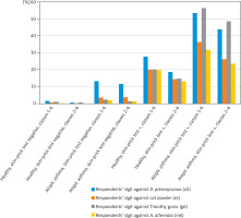
Relating to allergens of D. pteronyssinus, cat dander, and A. alternata, sIgE antibodies were more frequently detected in respondents with atopic asthma and a negative skin-prick test as compared to healthy respondents with a negative skin-prick test. Numerous statistically significant differences were identified (p < 0.005 to p < 0.001). Relating to allergens of D. pteronyssinus, cat dander, and timothy grass, sIgE antibodies were more frequently detected in respondents with atopic asthma and a weakly positive skin-prick test as compared to healthy respondents with a weakly positive skin-prick test. Numerous statistically significant differences were identified (p < 0.05 to p < 0.001) (Table 4, Figure 1).
Table 4
Number (percentage) of respondents with sIgE concentration ≥ 0.35 IU/ml (classes 1–6) or ≥ 0.7 IU/ml (classes 2–6) – healthy respondents and respondents with atopic asthma
Discussion
A broad ECAP survey proved the epidemiological significance of allergic rhinitis and asthma in Poland and the great diversity of allergy risk factors. The determination of specific IgE in respondents’ serum, a reliable method to evaluate allergic hypersensitivity, has been the continuation of ECAP. The aim of the study described in this article was to determine the relationship between the concentration of specific IgE antibodies in serum and types of asthma.
When comparing respondents with mild asthma to respondents with moderate asthma, no statistically significant differences were identified, although in respondents with moderate asthma, the percentages were frequently much greater than in respondents with mild asthma. The lack of the differences may result partially from the small number of analysed respondents, and because often the allergists did not define the type of asthma [12]. Similar values were obtained after excluding from the statistical analysis respondents with non-atopic asthma. No statistically significant differences were identified when comparing respondents with intermittent asthma to respondents with persistent asthma. Similar values were obtained after excluding from the statistical analysis respondents with non-atopic asthma. In a study by Matsui et al., allergen-specific IgE levels were associated with more severe asthma across a range of clinical and biologic markers [15]. In a study by Wickman et al., the presence of IgE antibodies seems not only to predict allergic diseases, but also relates to severity of such diseases, in particular to asthma [16]. In a study by Custovic et al., among children with wheezing, those in the complete mite sensitization trajectory had significantly higher risk of severe exacerbations [17]. In a study by Wang et al., sensitization to environmental allergens and total IgE correlated with increased healthcare and medication use, but not with wheeze symptoms [18]. In a study by Just et al. the following observations were made: a) children suffering from asthma with severe exacerbations and multiple allergies had more sensitisations to inhaled allergens and food allergens; b) children suffering from severe asthma with bronchial obstruction had significantly higher levels of all classes of immunoglobulin, except IgE; and c) children suffering from mild asthma did not show statistically significant features [19]. In a study by Manise et al., severe asthmatics had slightly increased total serum IgE compared with mild-to-moderate asthmatics without any difference in the sensitisation rate to common aeroallergens [20]. In a study by Haselkorn et al., severe or difficult-to-treat asthma in children and adolescents was characterized by high total serum IgE levels [21]. In a study by Satwani et al., serum total IgE levels were associated with severity of bronchial asthma [22]. In a study by Peat et al., subjects who had respiratory symptoms had higher total serum IgE levels than those who were lifelong asymptomatic [23]. In a study by Louis et al., variation in total serum IgE was weakly associated with asthma control but not with exacerbation [24].
Relating to allergens of D. pteronyssinus, cat dander, and A. alternata, sIgE antibodies were more frequently detected in respondents with atopic asthma and a negative skin-prick test as compared to healthy respondents with a negative skin-prick test. Relating to allergens of D. pteronyssinus, cat dander, and timothy grass, sIgE antibodies were more frequently detected in respondents with atopic asthma and a weakly positive skin-prick test as compared to healthy respondents with a weakly positive skin-prick test. Thus, relating to respondents with a negative or weakly positive skin-prick test, when sIgE antibodies against the same allergen are detected, occurrence of asthma is much more probable. If we suspect atopic asthma, determination of specific IgE antibodies is especially helpful in such patients. The results of this determination enhance the probability of a correct diagnosis. In a study by Simpson et al., the probability of wheeze and reduced lung respondents function increased with increasing specific IgE antibody levels, while there was no association between current wheeze and the size of skin test wheal [25]. In a study by Toppila-Salmi et al., polysensitization to more than one allergen type and the number of SPT-positive reactions was associated with asthma [26]. In a study by Chauveau et al., skin-prick test had a higher specificity for asthma and hay fever than sIgE, without difference for sensitivity [27]. In a study by Hourihane et al., peanut-specific IgE levels did not predict clinical severity, and skin-prick test weal size correlated weakly with severity [28]. In a study by Peat et al., total serum IgE was not more predictive of airway hyperresponsiveness than skin-prick tests [23]. In a study by Moore et al., the severe asthma group had less atopy by skin tests, but total IgE did not differentiate disease severity [29].
Conclusions
sIgE antibodies against any allergen are detected in 74.3% (classes 1 – 6)/69.0% (classes 2 – 6) of respondents with atopic asthma, but also in 9.9% (classes 1 – 6)/7.6% (classes 2 – 6) of healthy respondents. No statistically significant differences were identified when comparing sIgE antibodies of respondents with intermittent asthma to sIgE antibodies of respondents with persistent asthma. Regarding subjects with a negative or weakly positive skin test, when sIgE antibodies to the same allergen are detected, asthma is much more likely to occur.


