Introduction
Titanium dioxide (TiO2) consists of 1 titanium atom and 2 oxygen atoms; it is also known as titanium white or titania [1]. It naturally occurs in the form of 3 compounds: anatase, rutile, and brookite [2]. It is mainly used as an ingredient in the manufacturing process of paint and varnish products. TiO2 is widely used in many industries (Figure 1). Due to its properties, it increases strength, hardness, and reflectivity, thus reducing the risk of damage to the surface it covers. As a pigment, it is also used for the production of enamel, glaze, pharmaceuticals, paper, plastics, toothpastes, and cosmetics, including sunblock to reflect light from the skin [2, 3]. Currently, its annual consumption reaches 4 million tons, and it is one of the top 5 nanoparticles used in consumer products [4, 5]. TiO2 is also a commonly used food additive marked with the symbol E171, approved for use throughout the European Union [2]. Nevertheless, more recently, its potentially toxic properties and adverse health effects caused by exposure to the gastrointestinal tract have been increasingly discussed [4]. In 2019, at the request of the French Office for Food, Environment, and Occupational Health & Safety (ANSES), the French Government signed a decree suspending the use of E171 as a food additive in France in accordance with the precautionary principle. This decree entered into force on 1 January 2020 [5, 6].
There are some differences in TiO2 intake with food around the world. It is estimated that in the U.S. the daily supply of titanium oxide is on average about 1–2 mg/kg bw (body weight) by children under 10 years of age. In the UK, on the other hand, this supply fluctuates between 2–3 mg/kg bw at the same age; a similar trend is also observed in Germany. On the other hand, in adults a much lower intake of this component is observed, which ranges from 0.5 to 1 mg/kg bw. The escalated risk of children being exposed to TiO2 is attributed to increased consumption of products containing this compound [4]. The highest levels of total TiO2 are found in chewing gums and toothpastes, while lower levels are seen in bakery products such as cookies and muffins [2].
TiO2 absorption
TiO2 finds its way into the body mainly by air passages, from where it can spread to the bloodstream and, when administered intranasally, can reach the nervous system and more precisely the hippocampus in the brain [7, 8]. Research shows that TiO2 nanoparticles can penetrate the blood-brain barrier. Inhaled TiO2 NP molecules can contribute to the development of neurodegenerative diseases, causing damage to brain tissue [9]. In addition, they can change the permeability of the vesicular-capillary barrier and thus reach other organs in the body, such as the liver or kidneys [10]. Nano-TiO2 causes toxic effects in many organs demonstrating immunotoxicity, reproductive toxicity, and inhalation toxicity; however, nano particles can be excreted from the body through sweat or metabolic products [11]. Studies from recent years also show that TiO2 nanoparticles can get into the body over the digestive tract through the surrounding lymphoid tissue via medication, foods, drinks, and water containing the compound [12].
Effect of TiO2 on the intestines
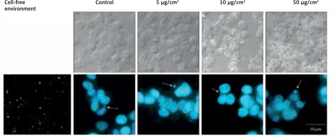
Many studies show the effect of titanium dioxide on the intestines. Ruiz et al. observed the effect of titanium dioxide administration on the course of enteritis in an animal study. They showed that exposure to TiO2 molecules can worsen the course of ongoing colitis. E171 can accumulate in intestinal epithelial cells, thereby leading to the accumulation of macrophages in this area. In addition, researchers have shown that patients with active ulcerative colitis (UC) have higher blood levels of titanium dioxide molecule, which proves that TiO2 can travel across the intestinal barrier. Depending on the dose taken, it can also affect the production of reactive oxygen species (ROS) [13]. Similar results were obtained by Proquin et al. They studied the effect of a food additive on genotoxicity in an in vitro study in Caco-2 and HCT-116 intestinal cell populations (Figure 2). The study showed that microparticles can cause ROS production. They also described chromosome damage caused by titanium dioxide exposure. Based on their observations the authors presented a hypothesis linking exposure to TiO2 particles with the occurrence of colorectal cancer [14].
Figure 2
Chromosome damage in HCT116 cells estimated by micronucleus assays (MN). Demonstrated 1.9-, 2.4-, and 3.6-fold of increase in cell cultures exposed to TiO2 in different doses [14]

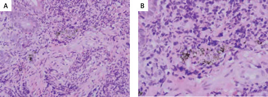
Gandamalla et al. found that the cytotoxicity of the molecule is dependent on the dose and its size. Small TiO2 particles cause the highest levels of toxicity [15]. Some of studies also prove that titanium dioxide can accelerate tumour growth and cause dysplastic changes in the epithelium of the large intestine in healthy people [16]. Tada-Oikawa et al. investigated the effect of titanium dioxide particles on the occurrence of inflammation in Caco-2 cells and THP-1 macrophages and their life span. They showed that the lifespan of THP-1 cells decreased after exposure to the structure of TiO2 anatase and TiO2 rutile after 24 h, while Caco-2 only after 72 h of exposure to all TiO2 structures. Inflammation was generated in both types of cells after exposure to TiO2 diameter of 50 nm in the amount of 50 µg/ml by the production of IL-1 in THP-1 and IL-8 in Caco-2 [17]. Yang et al. studied various types of titanium dioxide nanoparticles. They concluded that only T-NP (molecule smaller than 100 nm) at a dose of 50 and 100 µg/ml can cause Caco-2 cell apoptosis [18]. TiO2 can also have a negative effect on intestinal homeostasis. In an animal study, Bettini et al. demonstrated the formation of premalignant changes in the large intestine load at a dose of 10 mg/kg bw/day, and an increase in Th1 and Th17 in the spleen, which indicates ongoing inflammation [19]. In addition, Richter demonstrated in his in vitro study that exposure to TiO2 may interfere with metabolic homeostasis through limited glucose transport due to damage to intestinal epithelial cells. In contrast, Lactobacillus rhamnosus GG reduces the negative effect of the food additive [20]. Dudefoi, studying the effect of the molecule on human intestinal microbiota observed that the low concentration of TiO2 exposure to intestinal bacteria in vitro does not lead to changes in the intestinal microbiota. However, a reduction in Bacteroides ovatus and an increase in the amount of Clostridium cocleatum after exposure to 36 mg/l for 5 days on TiO2 particles has been shown. The authors indicate the need for research showing the effect of long-term use of TiO2 on intestinal microbiome [21]. Another study revealed that using TiO2 nanoparticles for 2–3 months can reduce strains such as Bifidobacterium and Lactobacillus. There is also scientific evidence confirming the relationship between TiO2 nanoparticles and the development of enteritis [22]. Particles can also accumulate in Peyer’s patches (Figure 3). Patients with IBD (more often with UC than with Crohn’s disease) found tuft deposits consisting of titanium dioxide, among others. It has been shown that older children have more of these deposits. The authors also suggest that the pigment detected in the terminal ileum may be associated with inflammation [23].
Figure 3
Biopsy results showed presence of the black pigment found in Peyer’s patches in the ileum. The pigment was located in the lymphoid follicle, outside the germinal centre [23]

In an animal model study, Blevins investigated the effect of TiO2 exposure on immune disorders. He showed that significant doses applied to rats (2617 mg/kg given for a total of 7 days or 29 400 mg/kg given for a total of 100 days) did not lead to inflammation in the gut or the circulatory system in food [24]. However, Mu et al. came to other conclusions. After 3 months of TiO2 administration to mice, they showed that nanoparticles, by reducing the amount of T cells (CD4) and macrophages, can interfere with the proper functioning of the immune system [22].
TiO2 and organs of the digestive system
The penetration capabilities of nano TiO2 show high potential for compound accumulation in many internal organs, indirectly leading to their damage. In animal models, intravenous nano-TiO2 administration has been shown to be associated with its accumulation in internal organs after 5 min, mainly in the liver (65%). Additionally, ICP-MS (inductively coupled plasma mass spectrometry) analysis showed that increasing doses of nano-TiO2 caused an increase in Ti4+ accumulation in the spleen and brain as well [11]. In the study of Chen et al., during 2 weeks of observation, mice were injected intraperitoneally with various doses of NP TiO2 (0, 324, 648, 972, 1296, 1944, or 2592 mg/kg body weight). Examination of the particle distribution showed that the accumulation of TiO2 NP (80 nm, 100 nm, anatase) was highest, successively, in the spleen and then in the liver, kidneys, and lungs [25]. The study suggest that NP TiO2 can be transported and deposited in other tissues or organs after intraperitoneal injection. However, the study used very high doses of exposure that could significantly affect the results obtained [12]. In addition, another study showed that exposure to high doses of TiO2 can lead to apoptosis of liver cells, necrosis and fibrosis, and renal glomerular swelling. Much of the data indicates that TiO2 may contribute to the growth of ROS. Nano-TiO2 enters the cell through the cell membrane and induces oxidative stress as a result of the imbalance between oxidants and antioxidants. As a consequence, it leads to lipid, protein, and DNA peroxidation, as well as cell apoptosis [11]. Jones et al. investigated the degree of absorption, excretion, or translocation of titanium dioxide in humans. TiO2 determination was performed using inductively coupled plasma mass spectrometry (ICP-MS) in the following fluids: blood, serum, urine, and culture medium. No relationship was found between the particle size and titanium dioxide and absorption in humans following oral administration of the compound. In addition, low absorption of titanium dioxide from the gastrointestinal tract after oral administration has been shown. There was no evident difference in absorption for any of the three particle sizes tested [26]. Some studies on acute oral toxicity of TiO2 have shown significant differences in clinical symptoms. In one study in animal models using higher doses of titanium dioxide (129.4 nm in H2O; 175, 550, 1750, or 5000 mg/kg; anatase 80/20/rutile; intervals of 48 h for 14 days) the observed effects were reversible. Oral use of lower doses of TiO2 particles (25, 80, and 155 nm; 5 g/kg bw) resulted in changes in biochemical parameters without systemic toxicity. The described deviations related to the level of alanine aminotransferase (ALT), aspartate aminotransferase (AST), lactate dehydrogenase (LDH), and blood urea nitrogen (BUN) [12]. In the case of studies on acute toxicity, other studies also confirm this relationship [27].
Impact of TiO2 on other organs
Many studies also indicate the undesirable effects of exposure to TiO2 in organs not belonging to the digestive system. Li et al. showed that NP TiO2 molecules are accumulated in significant amounts in the lungs, among others. They also observed that the molecules can damage DNA and induce inflammation as well as apoptosis of cells in the lungs [27]. Another study in animal models showed that TiO2 nanoparticles boosted the number of free radicals in the lungs and increased genotoxicity. In addition, it has been shown to be mutagenic, which can lead to cancer development [28]. Eye exposure of TiO2 is also unfavourable. Eom et al. conducted a study in animal models, in which they instilled TiO2 in rabbits’ right eyes, and then examined, among others, tear secretion and surface of the conjunctival goblet cells. They found that exposure to the ingredient may cause eye damage [29]. In many studies, TiO2 is presented as a component that causes genotoxicity and disorders in the proper functioning of the immune system [30]. In addition, it has been shown that there is increased production of ROS after ingredient exposure [31].
In the case of skin absorption, the test results are inconclusive [32]. Many of those studies have rated the absorption of TiO2 nanoparticles in healthy skin. NP TiO2 levels in the epidermis and dermis of individuals who used NP TiO2 on the skin were higher than NP TiO2 levels in the control group. Damaged and intact skin was subjected to UV radiation, deposited 102.35 ±4.20% and 102.84 ±5.67% Ti, respectively, on the surface and stratum corneum (SC), while only 0.19 ±0.15% and 0.39 ±0.39% was found in living epidermis and dermis. Substituting these results, the researchers concluded that TiO2 nanoparticles contained in the sunscreen remain in the highest SC layers, in both intact and damaged skin or skin exposed to simulated solar radiation [32, 33].
Conclusions
Titanium dioxide shows pro-inflammatory properties in the intestines, lungs, or the eyes and has the ability to accumulate in internal organs. It can also cause cyto- and genotoxicity. However, it should be noted that often the doses of TiO2 used in research are significantly higher than those that can enter the human body in everyday conditions. For this reason, the extent of absorption when exposed to the human body is still being questioned. In the future, it would be worth investigating the long-term exposure to the ingredient depending on its physicochemical structure. Due to the different routes of exposure to the component (oral, dermal, inhalation), the effects of the molecule on various systems should be thoroughly studied.


