Introduction
Leiomyomas are benign, well-circumscribed mesenchymal tumours [1]. While uterine leiomyomas are common, vulval leiomyomas are rare [2]. We report the case of a vulval leiomyoma in a multiparous woman, who was treated by excision procedure and developed acute postoperative delirium.
Material and methods
Consent
Written informed consent was obtained from the patient for her anonymised data including the images to be published. The procedures followed were in accordance with the Helsinki Declaration of 1975, revised in 2000.
Previous presentation
This manuscript has not been published or presented anywhere else before, nor is it being considered for publication anywhere else currently.
Ethical approval
The Institutional Review Board of our hospital does not require ethical approval for publication of case reports.
Case report
A 41-year-old multiparous woman presented with mass in the perineum of one year duration and discomfort in a sitting position since a month previously. There was no history of fever, local pain, pruritus vulvae, or discharge from the mass. Her bowel and bladder habits were normal. Her menstrual cycles were regular, with average flow, no dysmenorrhoea, and her last menstrual period was 18 days ago. She was para 2 with previous uneventful vaginal deliveries. Her last childbirth was 18 years previously, and she had been sterilised. Her history was uneventful and there was no family history of gynaecological malignancies or tumours. On examination, she was conscious, co-operative, and afebrile. Her pulse rate was 86/minute and blood pressure was 140/90 mm Hg. General and abdominal examination was unremarkable. On examination of the vulva, there was a mass of size 10 × 8 cm on the left side, involving the labium majus and minus, distorting the introitus, which was firm in consistency, with a small pus point on the muco-cutaneous junction (Fig. 1 A). On per speculum examination, the cervical os was obscured due to the mass. Per vaginal examination revealed the uterus to be anteverted, normal size, firm, mobile, with bilateral fornices free. A provisional diagnosis of Bartholin’s cyst was made, and she was further worked up.
Fig. 1
Preoperative images. (A) Vulval mass of size 10 × 8 cm on the left side, involving the labium majus (thick white arrow) and minus, distorting the introitus (thin white arrow). (B) Preoperative pelvic ultrasound showing a normal uterus (thin black arrow). (C) Transperineal ultrasound showing a solid lobulated mass with heterogenous echotexture (thick white arrow), suggestive of vulval fibroid

Her haemoglobin was 10.7 g/dl, total leucocyte count 10.07 × 10³/cumm, and liver and kidney function tests were within normal limits. Pelvic ultrasound was done, which showed a normal sized uterus and ovaries (Fig. 1 B). As the vulval mass was firm in consistency, transperineal ultrasound was performed, which revealed a solid lobulated mass with heterogenous echotexture, measuring 10 × 8 cm, with mild vascularity, in the left vulval region (Fig. 1 C). A preoperative diagnosis of vulval leiomyoma was made, and she was planned for excision under antibiotic cover.
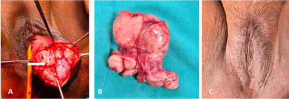
Under aseptic conditions and spinal anaesthesia, a vertical incision was made over the muco-cutaneous junction. The tumour was soft and fleshy, with well-defined planes (Fig. 2 A). Complete excision was performed with obliteration of the cavity. The excised tumour measured around 12 × 10 cm (Fig. 2 B).
Fig. 2
Intraoperative and postoperative images. (A) Intraoperatively, the tumour (thick white arrow) was soft and fleshy, with well-defined planes. (B) Completely excised specimen of vulval leiomyoma measuring 12 × 10 cm. (C) Good healing of local surgical site at discharge

Three hours postoperatively the patient was agitated, irritable, and trying to remove all intravenous (IV) lines. She was diagnosed with acute postoperative delirium and treated with an injection of haloperidol under a multidisciplinary team approach, to which she responded well. On postoperative day 1, her total leucocyte count was elevated, C-reactive protein was positive, and serum electrolytes were deranged (Table 1). She was treated with IV antibiotics, and her electrolyte imbalance was corrected. She recovered well and was discharged on the sixth postoperative day with good healing of the local surgical site (Fig. 2 C).
Table 1
Blood parameters
Histopathological gross examination revealed a single greyish white to greyish brown irregular lobulated mass measuring 12.0 × 10.0 × 7.0 cm with unremarkable outer surface. Serial slicing revealed a well encapsulated lesion. The cut surface was greyish white, solid, with no areas of necrosis or haemorrhage. No whorling pattern was seen. Few areas of cystic degeneration were noted. Microscopic examination showed a well circumscribed tumour comprising spindle shaped cells arranged in long intersecting fascicles. The tumour cells were elongated, spindle shaped, with vesicular nucleus and with inconspicuous nucleoli and moderate eosinophilic cytoplasm. Prominent myxohyaline changes and focal mucin pools were noted. Focal lymphoid aggregates, mast cells, and interspersed thin-walled blood vessels were noted. The number of mitoses were 0–1/10 high power field. There was no evidence of increased mitosis, necrosis, or marked nuclear atypia. On immunohistochemistry, the tumour cells were strongly and diffusely positive for SMA. The impression was leiomyoma of the vulval mass.
She was asymptomatic at 6-month follow-up and had no recurrence.
Discussion
Vulval leiomyomas are rare, accounting for 0.03% of all gynaecological neoplasms and 0.07% of all vulvar tumours [2–4]. A 2018 review identified 31 case reports of vulval leiomyomas across the globe [1]. Since then, a handful more have been described. Vulval leiomyoma usually occurs in the fourth and fifth decade of life, as in our case also. The size of the vulval leiomyomas previously described ranged 2–15 cm [1]. As the mass remains small during the initial period (10–20 years), patients are asymptomatic and may not present to the healthcare facility. As the mass increases in size, patients become symptomatic with difficulty in walking, sitting, having coitus, pain, pruritus, and erythema [1]. Compression of the bladder or rectum by vulval leiomyoma is infrequent.
Owing to the rarity, vulval leiomyoma is often misdiagnosed as Bartholin’s cyst preoperatively [5]. In our case, the firm nature of the tumour prompted further imaging, which revealed a solid lobulated mass suggestive of vulval leiomyoma. Both ultrasound and MRI have been used for preoperative characterisation [1, 6]. Excision is known to be therapeutic in both premenopausal and postmenopausal women. Recurrence may occur as the tumour appears to be hormone dependent; hence, long-term follow up is warranted.
Our case was unique because the patient also developed acute postoperative delirium following excision of the vulval leiomyoma. Delirium is defined as ‘acute brain dysfunction’, and symptoms include disorientation, perceptual disturbances, emotional dysregulation, or sleep disturbances [7]. Postoperative delirium usually happens in the recovery room and appears up to 5 days after surgery [8]. Its pathophysiology is poorly understood. Risk factors include the following: a) preoperative factors like patient anxiety, depression, advanced age, male sex, presence of comorbidities like diabetes mellitus and/or hypertension, dehydration, dementia, baseline cognitive dysfunction, history of transient ischaemic attacks, peripheral vascular disease, carotid and/or intracranial stenosis, atrial fibrillation, electrolyte abnormalities, sleep deprivation, alcohol abuse, smoking, and anticholinergic drugs; b) intraoperative factors like site of surgery (abdominal, hip, and cardiothoracic), open surgery, hypotension, shock, hypothermia or hyperthermia, arrhythmias, increased surgical duration, haemorrhage, and blood transfusion; c) postoperative factors like prolonged intubation, pain, liver/kidney failure, anaemia, hypoxaemia, hypoalbuminaemia, benzodiazepine withdrawal, and sleep cycle disturbances [9, 10].
To the best of our knowledge, acute postoperative delirium has not been previously reported after the excision of a vulval leiomyoma. Probably, in our case, prior patient anxiety along with open surgical excision of a large mass from a relatively small vulval area and electrolyte imbalance was causative. Anaesthetic drugs seem unlikely to be causative or a contributory factor in our case because general anaesthesia was not administered. Management of postoperative delirium is supportive and focusses on the precipitating factors. Pharmacological therapy is used where there is a safety risk to the patient. In our case, she responded well to haloperidol, dyselectrolytaemia correction, and supportive measures.


