eISSN: 1731-2531
ISSN: 1642-5758
Anaesthesiology Intensive Therapy
Current issue Archive Manuscripts accepted About the journal Supplements Editorial board Reviewers Subscription Contact Instructions for authors Publication charge Ethical standards and procedures
Editorial System
Submit your Manuscript
SCImago Journal & Country Rank
3/2024
vol. 56
 
Share:
Share:
Original article

Correlations between preoperative fluid status assessed by bioimpedance analysis and hypotension during anaesthesia induction

Ianis Siriopol
1, 2
,
Ioana Grigoras
1, 2
,
Daniel Rusu
2
,
Raluca Popa
3
,
Irina Ristescu
1, 2
,
Mehmet Kanbay
4
,
Dimitrie Siriopol
3, 5

  1. “Grigore T. Popa” University of Medicine and Pharmacy, Iași, Romania
  2. Department of Anaesthesia and Intensive Care, Regional Institute of Oncology, Iași, Romania
  3. Department of Nephrology, “Sf. Ioan cel Nou” County Hospital, Suceava, Romania
  4. Division of Nephrology, Department of Internal Medicine, Koc University School of Medicine, Istanbul, Turkey
  5. “Stefan cel Mare” University, Suceava, Romania
Anaesthesiol Intensive Ther 2024; 56, 3: 177–184
Online publish date: 2024/08/30
Article files
- Correlations.pdf  [0.20 MB]
Get citation
 
PlumX metrics:
 

Hypotension during general anaesthesia lasting for even short periods of time can have deleterious effects in the postoperative period [1]. Multi-ple studies have demonstrated the relationship between intraoperative hypotension (IOH) and increased postoperative morbidity and mortality secondary to IOH-induced organ injury (myocardial lesions, stroke, kidney injury, splanchnic hypoperfusion) in surgical patients [1, 2]. Identifying patients at high risk of haemodynamic instability during anaesthesia induction can allow the anaesthesiologist to modify the anaesthetic dosing or administration rate, pre-emptive fluid administration or initial vasoactive medication use [3]. The stability of blood pressure during induction of anaesthesia is of major importance as hypotension during this period may lead to decreased tissue perfusion and organ injury, with an adverse impact on patient outcome [4, 5]. The probability of postinduction hypotension (PIH) increases with age, body mass index, increased blood pressure at the time of operating room arrival, diabetes mellitus type II and high ASA scores [6, 7], most of which are unmodifiable risk factors for PIH. Latent volume depletion is a modifiable risk factor of high clinical relevance.

Correct assessment of fluid status is a difficult task in clinical practice. Hypovolaemia is presumed to be a common factor of PIH, despite worldwide improvement in preoperative volume optimization, avoiding unnecessary fasting and bowel preparation [7]. The utility of non-invasive ultrasound assessment by anaesthesiologists could be important in the assessment of haemodynamic status [8]. Although several studies have investigated the use of preoperative inferior vena cava ultrasound examination, its use for PIH prediction is heterogenous both in methodo-logy and in results, with a large difference between observed sensitivities and specificities [9]. Therefore, preoperative identification of latent hypovolaemia can prompt the anaesthetist to ini-tiate fluid admi-nistration before induction of anaesthesia.

In patients undergoing chronic haemodialysis, bioimpedance-based methods are frequently used to measure body composition [10, 11]. Bioimpe-dance analysis (BIA) is used to assess body composition and estimate total and extracellular water volumes based on the tissue’s capacity to conduct electrical impulses [12]. Bioimpedance spectroscopy (BIS) is a safe, simple, fast and unique bioimpedance approach that is frequently used in clinical practice [13]. As compared with other methods, which are costly, unnecessarily invasive in most elective surgical procedures, with high complication rates (pulmonary arterial catheter, transpulmonary haemodilution technique, etc.) [14] or highly heterogenous in estimation [9], BIS objectively and easily assesses the preoperative fluid status, and may indicate fluid depletion correction, avoiding hypotension at anaes-thesia induction.

The study’s main purpose was to determine the correlation between the preoperative BIA assessed fluid status and PIH.

Methods

This study was performed on the same cohort of patients as a previously published prospective observational single centre study which investigated the relationship between arterial stiffness and PIH. All the details of the study design, inclusion and exclusion criteria have been previously published [15].

Bioimpedance measurement

Bioimpedance measurements were performed with a commercial device, the Body Composition Monitor (BCM; Fresenius Medical Care, Bad Homburg, Germany), a portable whole-body multifrequency bioimpedance analysis device. The BCM measures body resistance and reactance to electrical currents of 50 discrete frequencies, ranging from 5 to 1000 kHz [16]. It can assess total body water (TBW) and extracellular water (ECW) [8]. The Chamney physiological tissue model, which considers the person’s weight, height, and gender, is used to provide values for TBW and ECW [17].

The BCM displays absolute fluid overload (AFO), the difference between normal (expected) and actual (measured) ECW (expressed in litres) and the relative fluid overload (RFO), calculated as absolute fluid overload/extracellular water ratio (AFO/ECW) expressed in percentages. A negative AFO indicates the patient’s underhydration, while a positive one indicates overhydration. Based on RFO values, we classified the hydration status into three categories: dehydrated (RFO < –10%), normohydrated (–10% ≤ RFO ≤ 15%), and hyperhydrated (RFO > 15%).

The bioimpedance measurements were performed by a trained operator according to the manu-facturer’s recommendations. The measurement was performed 2 hours before the surgery. After explaining the procedure, blood pressure, body height, and weight were measured and the patient placed in the supine position. After cleaning the skin with alcohol solution, four dedicated skin electrodes were placed on the right wrist and ankle. To minimize electrical disturbances at the time of measurement, contacts between the patient and metallic bed parts were excluded. The patient’s weight and height were entered into the device, and measurements were recorded. Successful completion of the measurement was checked in real time on the device using a signal quality index > 85% and a Cole-Cole Gaussian curve. Measured and derived parameters were displayed within 2 minutes.

Induction of anaesthesia protocol and PIH

As mentioned in the published data [15], we protocolized pre-anaesthetic evaluation and anaesthesia induction. We defined PIH as the blood pressure decrease occurring during the first 10 minutes after induction of anaesthesia and orotracheal intubation before surgical incision and used the same definition for hypotension as in the previously published study [15]. Baseline blood pressure was the one measured 2 hours before induction of anaesthesia, when BIA was performed. During the intervention, non-invasive arterial blood pressure was assessed every minute during the first 10 minutes after the induction of anaesthesia.

Statistical analysis

All analyses were performed using Stata SE software, version 13 (Stata Statistical Software: Release 12. College Station, TX: Stata Corp LP). Continuous data are presented as mean ± standard deviation (SD), median with interquartile range (IQR), and categorical data as number and percent frequency. The between-group comparisons were performed using the χ2 or Fisher test, the Mann-Whitney U test or the independent t-test, as appropriate. The distribution of the variables was assessed using the Shapiro-Wilk test.

Logistic and linear regression analyses were used to assess the relationships between different variables derived from the bioimpedance analysis (TBW, ECW, intracellular water [ICW], AFO, RFO and ECW to ICW ratio) and PIH incidence and duration. For the multiva-riable analysis, as in the previous analysis [15], we selected a priori, based on existing knowledge and literature, the following confounders: age, gender, ASA physical status, comorbidities (coronary artery disease, blood hypertension, diabetes mellitus and malignancy) and medication (β-blocker and angiotensin II receptor blockers/angiotensin converting enzyme inhibitors use). When needed, we performed bootstrapping validation to avoid the problem of overfitting owing to the low number outcomes (PIH at different thresholds). A 2-tailed P < 0.05 was considered statistically significant.

Results

Study population characteristics

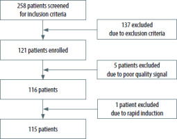
Out of the 258 patients screened for inclusion criteria, 115 patients were included in the final ana-lysis (Figure 1).

FIGURE 1

Flow chart of the study

/f/fulltexts/AIT/54712/AIT-56-54712-g001_min.jpg

The baseline characteristics of the study population were previously presented [12]. The mean age was 58.1 years and 69.6% were female (Table 1). More than 60% of the study population had the dia-gnosis of cancer.

TABLE 1

Baseline characteristics of the study population (N = 115)

Variables
Age (years)58.1 ± 11.4
Male, n (%)35 (30.4)
BMI, kg m–227.5 ± 4.2
MBP (mmHg)104.0 ± 10.5
SBP (mmHg)139.5 ± 17.5
DBP (mmHg)84.5 ± 10.24
Anthropometric data
     Weight (kg)75.7 ± 15.6
Bioimpedance measurements
     TBW (L)35.1 (31.5–40.8)
ECW (L)15.7 (14.0–18.7)
ICW (L)19.3 (17.1–22.6)
AFO (L)–0.1 (–1.0–0.7)
RFO (%)–0.6 ± 8.3

[i] Data are expressed as mean ± standard deviation, median (interquartile range), or as number (percentage).

[ii] AFO – absolute fluid overload, BMI – body mass index, DBP – diastolic blood pressure, ECW – extracellular water, ICW – intracellular water, MBP – mean blood pressure, RFO – rela-tive fluid overload, SBP – systolic blood pressure, TBW – total body water

The incidence of PIH, defined as a decrease in systolic (SBP) and mean blood pressure (MBP), or a as a relative decrease of SBP and MBP from baseline, is presented in Table 2.

TABLE 2

Incidence of postinduction hypotension (PIH) using different thresholds

PIH thresholdsn (%)
Absolute SBP (mmHg)
     < 100100 (86.9)
< 9066 (57.4)
< 8039 (33.9)
< 7017 (14.8)
Relative SBP thresholds (%)
     > 10115 (100)
> 20114 (99.1)
> 30101 (87.8)
> 4063 (54.8)
Absolute MBP (mmHg)
     < 7086 (74.8)
< 6040 (34.8)
< 5011 (9.6)
< 400 (0)
Relative MBP thresholds (%)
     > 10115 (100)
> 20113 (98.3)
> 3097 (84.4)
> 4059 (51.3)

[i] MBP – mean blood pressure, SBP – systolic blood pressure

BIA assessed fluid status and incidence of PIH

In the univariable analysis, TBW was associated with the development of PIH when defined as an absolute SBP decrease < 80 mmHg and as a relative MAP decrease of > 20% from baseline (Table 3). In the multivariable analysis, only the association with an absolute SBP decrease < 80 mmHg remained statisti-cally sig-nificant (OR = 0.91, 95% CI: 0.83–0.99) (Table 3).

TABLE 3

The relationship between total body water (L) and postinduction hypotension (PIH) using different thresholds

PIH thresholdsOR* (95% CI)P-valueAdjusted# OR* (95% CI)P-value
Absolute SBP (mmHg)
     < 1000.98 (0.91 to 1.05)0.490.92 (0.81 to 1.03)0.15
< 900.95 (0.90 to 1.00)0.050.95 (0.88 to 1.02)0.17
< 800.94 (0.88 to 0.99)0.040.91 (0.83 to 0.99)0.03
< 700.95 (0.87 to 1.03)0.190.97 (0.86 to 1.09)0.62
Relative SBP thresholds (%)
     > 10NANANANA
> 200.89 (0.74 to 1.09)0.260.92 (0.62 to 1.37)0.67
> 300.97 (0.91 to 1.04)0.370.90 (0.79 to 1.02)0.11
> 400.97 (0.92 to 1.02)0.230.96 (0.88 to 1.04)0.33
Absolute MBP (mmHg)
     < 700.97 (0.92 to 1.03)0.330.99 (0.92 to 1.09)0.99
< 600.95 (0.89 to 1.01)0.080.95 (0.87 to 1.04)0.28
< 500.95 (0.86 to 1.05)0.291.04 (0.88 to 1.23)0.64
< 40NANANANA
Relative MBP thresholds (%)
     > 10NANANANA
> 200.86 (0.75 to 0.99)0.040.76 (0.55 to 1.07)0.12
> 300.96 (0.91 to 1.03)0.241.01 (0.91 to 1.11)0.91
> 400.98 (0.93 to 1.03)0.410.99 (0.91 to 1.07)0.79

# Adjusted for age, gender, ASA score, comorbidities and chronic medication.

* ORs are per 1 L increase in the absolute fluid overload.

CI – confidence interval, MBP – mean blood pressure, NA – not applicable, OR – odds ratio, SBP – systolic blood pressure

Similar results were obtained for ICW. It was asso-ciated with the development of PIH when defined as an absolute SBP decrease < 80 mmHg and as a relative MAP decrease of > 20% from baseline (Table 4). In the multivariable analysis, only the association with an absolute SBP decrease < 80 mmHg remained statistically significant (OR = 0.85, 95% CI: 0.74–0.99) (Table 4).

TABLE 4

The relationship between intracellular water (L) and postinduction hypotension (PIH) using different thresholds

PIH thresholdsOR* (95% CI)P-valueAdjusted# OR* (95% CI)P-value
Absolute SBP (mmHg)
     < 1000.97 (0.86 to 1.09)0.650.91 (0.75 to 1.09)0.31
< 900.93 (0.85 to 1.01)0.090.95 (0.83 to 1.08)0.42
< 800.89 (0.80 to 0.99)0.030.85 (0.74 to 0.99)0.03
< 700.92 (0.81 to 1.06)0.250.99 (0.82 to 1.19)0.88
Relative SBP thresholds (%)
     > 10NANANANA
> 200.79 (0.55 to 1.17)0.240.81 (0.39 to 1.68)0.57
> 300.93 (0.82 to 1.05)0.210.83 (0.66 to 1.04)0.10
> 400.94 (0.86 to 1.03)0.180.95 (0.83 to 1.09)0.49
Absolute MBP (mmHg)
     < 700.96 (0.87 to 1.06)0.411.02 (0.88 to 1.17)0.85
< 600.92 (0.84 to 1.01)0.090.94 (0.82 to 1.09)0.44
< 500.91 (0.77 to 1.08)0.281.08 (0.84 to 1.39)0.56
< 40NANANANA
Relative MBP thresholds (%)
     > 10NANANANA
> 200.72 (0.53 to 0.99)0.040.61 (0.33 to 1.11)0.11
> 300.92 (0.822 to 1.03)0.130.97 (0.82 to 1.15)0.74
> 400.94 (0.86 to 1.02)0.160.95 (0.83 to 1.09)0.49

* ORs are per 1 L increase in the absolute fluid overload.

# Adjusted for age, gender, ASA score, comorbidities and chronic medication.

CI – confidence interval, MBP – mean blood pressure, NA – not applicable, OR – odds ratio, SBP – systolic blood pressure

There was no association between ECW, AFO, RFO, and PIH incidence in the univariable and multivariable analysis, as shown in Supplementary Tables 1, 2 and 3, respectively.

Supplementary Tables

BIA assessed fluid status and duration of PIH

There was no correlation between any of the BIA-derived parameters of fluid status and the duration of PIH (see Tables 5 and 6 and Supplementary Tables 4, 5 and 6).

TABLE 5

The relationship between total body water and duration of postinduction hypotension (PIH) using different thresholds

PIH thresholdsβ coefficient* (95% CI)P-valueAdjusted# β coefficient* (95% CI)P-value
Absolute SBP (mmHg)
     < 100–0.08 (–0.17 to 0.01)0.07–0.13 (–0.27 to 0.01)0.05
< 90–0.05 (–0.12 to 0.02)0.16–0.06 (–0.17 to 0.04)0.24
< 80–0.02 (–0.06 to 0.01)0.18–0.05 (–0.10 to 0.01)0.06
< 70–0.01 (–0.02 to 0.11)0.52–0.01 (–0.04 to 0.01)0.23
Relative SBP thresholds (%)
     > 10–0.04 (–0.09 to 0.02)0.18–0.06 (–0.13 to 0.02)0.16
> 20–0.05 (–0.11 to 0.02)0.19–0.03 (–0.13 to 0.07)0.59
> 30–0.04 (–0.14 to 0.05)0.35–0.02 (–0.15 to 0.11)0.73
> 40–0.01 (–0.08 to 0.07)0.920.02 (–0.08 to 0.12)0.64
Absolute MBP (mmHg)
     < 70–0.05 (–0.13 to 0.02)0.18–0.05 (–0.16 to 0.06)0.36
< 60–0.02 (–0.06 to 0.02)0.29–0.02 (–0.07 to 0.03)0.48
< 50–0.01 (–0.01 to 0.01)0.710.01 (–0.01 to 0.02)0.44
< 40NANANANA
Relative MBP thresholds (%)
     > 10NANANANA
> 20–0.06 (–0.13 to 0.02)0.12–0.02 (–0.13 to 0.08)0.66
> 30–0.05 (–0.14 to 0.04)0.300.02 (–0.12 to 0.15)0.79
> 40–0.01 (–0.8 to 0.05)0.710.01 (–0.08 to 0.11)0.79

* β coefficients are per 1 L increase in the total body water.

# Adjusted for age, gender, ASA score, comorbidities and chronic medication.

CI – confidence interval, MBP – mean blood pressure, NA – not applicable, SBP – systolic blood pressure

TABLE 6

The relationship between intracellular water and duration of postinduction hypotension (PIH) using different thresholds

PIH thresholdsβ coefficient* (95% CI)P-valueAdjusted# β coefficient* (95% CI)P-value
Absolute SBP (mmHg)
     < 100–0.13 (–0.29 to 0.03)0.09–0.19 (–0.41 to 0.04)0.09
< 90–0.08 (–0.20 to 0.04)0.20–0.07 (–0.25 to 0.11)0.43
< 80–0.04 (–0.10 to 0.02)0.18–0.07 (–0.16 to 0.01)0.09
< 70–0.01 (–0.03 to 0.02)0.74–0.01 (–0.05 to 0.03)0.49
Relative SBP thresholds (%)
     > 10–0.06 (–0.15 to 0.03)0.21–0.08 (–0.21 to 0.05)0.23
> 20–0.09 (–0.22 to 0.03)0.14–0.05 (–0.22 to 0.13)0.61
> 30–0.10 (–0.27 to 0.06)0.21–0.03 (–0.26 to 0.19)0.76
> 40–0.04 (–0.17 to 0.09)0.570.03 (–0.14 to 0.19)0.72
Absolute MBP (mmHg)
     < 70–0.09 (–0.22 to 0.05)0.20–0.06 (–0.24 to 0.12)0.51
< 60–0.04 (–0.11 to 0.02)0.21–0.03 (–0.12 to 0.06)0.45
< 50–0.01 (–0.03 to 0.01)0.520.01 (–0.02 to 0.04)0.58
< 40NANANANA
Relative MBP thresholds (%)
     > 10NANANANA
> 20–0.12 (–0.23 to 0.01)0.07–0.06 (–0.24 to 0.11)0.48
> 30–0.12 (–0.28 to 0.05)0.160.01 (–0.22 to 0.23)0.99
> 40–0.06 (–0.18 to 0.05)0.29–0.02 (–0.17 to 0.14)0.83

* β coefficients are per 1 L increase in the intracellular water.

# Adjusted for age, gender, ASA score, comorbidities and chronic medication.

CI – confidence interval, MBP – mean blood pressure, NA – not applicable, SBP – systolic blood pressure

Discussion

To our knowledge, this is the first study that has investigated the relationship between fluid status assessed using BIA and PIH. We found that TBW and its ICW component were the only two BIA-derived parameters associated with the development of PIH.

BIA is a simple technique that provides an objective assessment of fluid status with numerical results. It is helpful even in the case of patients with subclinical abnormalities. BIA was validated by isotope dilution methods [11], the gold standard for assessing body fluid composition, and is commonly used to guide fluid removal in dialysis patients [16, 18]. BIA defines the hydration status of an individual based on normal extracellular volume and body composition [19]. It performs well for healthy subjects and chronic patients with major and subtle water distribution disturbances [20]. BIA measures ECW but cannot discriminate between extravascular and intravascular volume [21]. Steinwandel et al. [22] evaluated whether intravascular volume status in haemodialysis patients measured by inferior vena cava ultrasound correlates with BIA values. Their results showed agreement between the two fluid assessment methods, concluding that both can determine the risk of intradialytic hypotension [22]. We used the cut-off BIA value for defining normo-, over- and dehydrated patients, as recommended for chronic patients undergoing haemodialysis. Other authors used the same values for perioperative fluid status assessment [23].

The ability to assess patient fluid status is an essential part of perioperative care. There is a need to identify easily available variables that can help clinicians recognize a modifiable risk factor for developing hemodynamic instability during sur--gery [24]. Hypovolaemia is probably the most important factor associated with PIH [7]. In the case of overt hypovolaemia, anaesthesia induction protocols are adapted to avoid or minimize PIH. Subtle, subclinical dehydration escapes clinical exa-mination. Based on the assumption of preoperative dehydration, administering a variable amount of intravenous fluid before anaesthesia induction is frequently encountered in clinical practice, even though increased intraoperative fluid balance is associated with increased postoperative morbidity and mortality [25, 26]. Preoperative BIA examination is a sensitive method that allows objective assessment of patient fluid status, being able to detect subclinical changes.

Using a protocol for anaesthesia induction and monitoring, we aimed to exclude induction agents as risk factors for PIH.

As previously reported for the same study group population, arterial stiffness identifies patients at risk of developing severe hypotension defined as an SBP or MBP decrease of more than 40% from baseline [15]. Also, arterial stiffness, evaluated by carotid-femoral pulse wave velocity, is significantly associated with the duration of PIH, defined as above [15]. This means that both factors, fluid status and arterial stiffness, could influence PIH. The exact mechanism by which these physiopathological pathways interact needs further investigation in the context of personalized medicine.

Administering large amounts of fluids during the induction period could result in hypervolaemia, which is harmful to vascular barrier competence [27]. Infusing fluids in a primarily normovolaemic circulation may lead to a breakdown of the endothelial surface layer [28], altering patient outcome. A more physiological strategy to prevent PIH is the use of vasopressor to re-establish blood pressure, fluids being a second-line strategy.

There is a high variability of PIH (5-99%), mainly due to the absence of an accepted definition of IOH [29]. Jor et al. [7] identified an incidence of PIH of 36.5%. Our results show a much higher incidence of PIH when using the same definition of PIH, 84.4%. The important difference between the PIH incidence is probably due to the frequency of blood pressure determination after induction, as we determined the blood pressure every minute and Jor et al. determined it only twice, at 5 and 10 minutes after induction.

Considering that, in the literature, there is strong evidence to support the relationship between IOH and increased postoperative morbidity and mortality after non-cardiac surgery [2, 30], we did not intend to quantify PIH effects on clinical outcome variables, as our study was designed to evaluate whether preoperative BIA assessment could identify patients at risk of PIH. Furthermore, although seve-ral studies have reported an association between intraoperative mean arterial blood pressure, especially relative decreases, with worse outcomes [3134], we did not identify any relationship between BIA-derived parameters and MBP.

Strengths and limitations

Our study excluded patients with comorbidities recognized as risk factors for PIH. By protocolizing patient preoperative preparation, induction of anaes-thesia and monitoring during anaesthesia induction, we excluded confounders. We evaluated the decrease of blood pressure during induction of anaesthesia using multiple definitions for PIH for both SBP and MBP. Also, baseline blood pressure was measured outside the operating theatre. This is important because most studies evaluating IOH use the first blood pressure measured in the operating room as baseline blood pressure. This may reflect transient hypertension at arrival in the operating room, thus overestimating the severity of PIH.

There are multiple limitations of our study. We included a relatively small sample size of elective surgical patients from a single-centre unit, with a high incidence of known cancer disease. Our findings regarding the BIA values measured in this group of patients might not apply to other settings because all patients underwent surgery in a single university tertiary medical centre, treating the patients according to local clinical standards. Our findings need to be verified by larger or multi-centric studies.

Conclusions

Our study shows that in elective surgery, BIA could detect subtle, subclinical fluid parameters that are associated with PIH.

Acknowledgements

Assistance with the article

none.

Financial support and sponsorship

none.

Conflicts of interest

none.

Presentation

none.

Ethics approval

The study was approved by the Institutional Review Board of the “Grigore T. Popa” University of Medicine and Pharmacy, Iasi, with approval number: 140724/2016.

References

1 

Walsh M, Devereaux PJ, Garg AX, Kurz A, Turan A, Rodseth RN, et al. Relationship between intraoperative mean arterial pressure and clinical outcomes after noncardiac surgery: toward an empirical definition of hypotension. Anesthesiology 2013; 119: 507-515. DOI: 10.1097/ALN.0b013e3182a10e26.

2 

Bijker JB, Persoon S, Peelen LM, Moons KGM, Kalkman CJ, Kapelle LJ, et al. Intraoperative hypotension and perioperative ischemic stroke after general surgery, a nested case-control study. Anesthesiology 2012; 116: 658-664. DOI: 10.1097/ALN.0b013e3182472320.

3 

Subramaniam B, Subramaniam K. Cautious optimism: can preoperative ultrasound predict postinduction hypotension? Anesthesiology 2016; 124: 526-527. DOI: 10.1097/ALN.0000000000001003.

4 

Joo HS, Salasidis GC, Kataoka MT, Mazer CD, Naik VN, Chen RB, et al. Comparison of bolus remifentanil versus bolus fentanyl for induction of anesthesia and tracheal intubation in patients with cardiac disease. J Cardiothorac Vasc Anesth 2004; 18: 263-268. DOI: 10.1053/j.jvca.2004.03.003.

5 

Bouillon TW, Bruhn J, Radulescu L, Andresen C, Shafer TJ, Cohane C, et al. Pharmacodynamic interaction between propofol and remifen-tanil regarding hypnosis, tolerance of laryngoscopy, bispectral index, and electroencephalographic approximate entropy. Anesthesiology 2004; 100: 1353-1372. DOI: 10.1097/00000542-200406000-00006.

6 

Reich DL, Hossain S, Krol M, Baez B, Patel P, Bernstein A, et al. Predictors of hypotension after induction of general anesthesia. Anesth Analg 2005; 101: 622-628. DOI: 10.1213/01.ANE.0000175214.38450.91.

7 

Jor O, Maca J, Koutna J, Gemrotova M, Vymazal T, Litschmannova M, et al. Hypotension after induction of general anesthesia: occurrence, risk factors, and therapy. A prospective multicentre observational study. J Anesth 2018; 32: 673-680. DOI: 10.1007/s00540-018-2532-6.

8 

Ferreira Albuquerque Costa NT, Gomar Sancho C. Perioperative ultrasound applied to diagnosis and decision making in anesthesia. Minerva Anestesiol 2018; 84: 94-107. DOI: 10.23736/S0375-9393.17.12178-4.

9 

Chowdhury SR, Datta PK, Maitra S, Rawat D, Baidya DK, Roy A, et al.The use of preoperative inferior vena cava ultrasound to predict anaesthesia-induced hypotension: a systematic review. Anaesthesiol Intensive Ther 2023; 55: 18-31. DOI: 10.5114/ait.2023.125310.

10 

Earthman C, Traughber D, Dobratz J, Howell W. Bioimpedance spectroscopy for clinical assessment of fluid distribution and body cell mass. Nutr Clin Pract 2007; 27: 389-405. DOI: 10.1177/0115426507022004389.

11 

Wabel P, Chamney P, Moissl U, Jirka T. Importance of whole-body bioimpedance spectroscopy for the management of fluid balance. Blood Purif 2009; 27: 75-80. DOI: 10.1159/000167013.

12 

Ciumanghel AI, Grigoras I, Siriopol D, Blaj M, Rusu DM, Grigorasi GR, et al. Bio-electrical impedance analysis for perioperative fluid evaluation in open major abdominal surgery. J Clin Monit Comput 2020; 34: 421-432. DOI: 10.1007/s10877-019-00334-8.

13 

Dewitte A, Carles P, Joannès-Boyau O, Fleureau C, Roze H, Combe C, et al. Bioelectrical impedance spectroscopy to estimate fluid balance in critically ill patients. J Clin Monit Comput 2016; 30: 227-233. DOI: 10.1007/s10877-015-9706-7.

14 

Vincent JL, Pelosi P, Pearse R, Payen D, Perel A, Hoeft A, et al. Peri-operative cardiovascular monitoring of high-risk patients: a consensus of 12. Crit Care 2015; 19: 224. doi: 10.1186/s13054-015-0932-7.

15 

Siriopol I, Grigoras I, Siriopol D, Ciumanghel A, Rusu D, Blaj M, et al. Arterial stiffness as a risk factor for hypotension during induction of general anesthesia. Arch Med Sci 2021. DOI: 10.5114/aoms/132836.

16 

Onofriescu M, Hogas S, Voroneanu L, Apetrii M, Nistor I, Kanbay M, et al. Bioimpedance-guided fluid management in maintenance hemodialysis: A pilot randomized controlled trial. Am J Kidney Dis 2014; 64: 111-118. DOI: 10.1053/j.ajkd.2014.01.420.

17 

Chamney PW, Wabel P, Moissl UM, Müller MJ, Bosy-Westphal A, Korth O, et al. A whole-body model to distinguish excess fluid from the hydration of major body tissues. Am J Clin Nutr 2007; 85: 80-89. DOI: 10.1093/ajcn/85.1.80.

18 

Moissl U, Arias-Guillén M, Wabel P, Fontseré N, Carrera M, Campistol JM, et al. Bioimpedance-guided fluid management in hemodialysis patients. Clin J Am Soc Nephrol 2013; 8: 1575-1582. DOI: 10.2215/CJN.12411212.

19 

Siriopol I, Siriopol D, Voroneanu L, Covic A. Predictive abilities of baseline measurements of fluid overload, assessed by bioimpedance spectroscopy and serum N-terminal pro-B-type natriuretic peptide, for mortality in hemodialysis patients. Arch Med Sci 2017; 13: 1121-1129. DOI: 10.5114/aoms.2017.68993.

20 

Bioelectrical impedance analysis in body composition measurement: National Institutes of Health Technology Assessment Conference statement. Am J Clin Nutr 1996; 64 (3 Suppl): 524S-532S. DOI: 10.1093/ajcn/64.3.524S.

21 

Jaeger JQ, Mehta RL. Dry weight and body composition in hemo-dialysis: a proposal for an index of fluid removal. Semin Dial 1999; 12: 164-174. DOI: 10.1046/j.1525-139X.1999.99011.x.

22 

Steinwandel U, Gibson N, Towell-Barnard A, Parsons R, Rippey J, Rosman J. Does the intravascular volume status in haemodialysis patients measured by inferior vena cava ultrasound correlate with bioimpedance spectroscopy? J Clin Nurs 2019; 28: 2135-2146. DOI: 10.1111/jocn.14804.

23 

Ernstbrunner M, Kostner L, Kimberger O, Wabel P, Säemann M, Markstaller K, et al. Bioimpedance spectroscopy for assessment of volume status in patients before and after general anaesthesia. PLoS One 2014; 9: e111139. DOI: 10.1371/journal.pone.0111139.

24 

Szabó M, Bozó A, Darvas K, Horváth A, Iványi ZD. Role of inferior vena cava collapsibility index in the prediction of hypotension associated with general anesthesia: an observational study. BMC Anesthesiol 2019; 19: 139. DOI: 10.1186/s12871-019-0809-4.

25 

Lobo DN, Bostock KA, Neal KR, Perkins AC, Rowlands BJ, Allison SP. Effect of salt and water balance on recovery of gastrointestinal function after elective colonic resection: a randomised controlled trial. Lancet 2002; 359: 1812-1818. DOI: 10.1016/S0140-6736(02)08711-1.

26 

Brandstrup B, Tønnesen H, Beier-Holgersen R, Hjortsø E, Ørding H, Lindorff-Larsen K, et al. Effects of intravenous fluid restriction on postoperative complications: comparison of two perioperative fluid regimens–a randomized assessor-blinded multicenter trial. Ann Surg 2003; 238: 641-648. DOI: 10.1097/01.sla.0000094387.50865.23.

27 

Jacob M, Chappell D. Effects of perioperative fasting on haemodynamics and intravascular volumes. Best Pract Res Clin Anaesthesiol 2012; 26: 421-430. DOI: 10.1016/j.bpa.2012.10.002.

28 

Rehm M, Haller M, Orth V, Kreimeier U, Jacob M, Dressel H, et al. Changes in blood volume and hematocrit during acute preoperative volume loading with 5% albumin or 6% hetastarch solutions in patients before radical hysterectomy. Anesthesiology 2001; 95: 849-856. DOI: 10.1097/00000542-200110000-00011.

29 

Bijker JB, Van Klei WA, Kappen TH, van Wolfswinkel L, Moons KGM, Kalkman CJ. Incidence of intraoperative hypotension as a function of the chosen definition: Literature definitions applied to a retrospective cohort using automated data collection. Anesthesiology 2007; 107: 213-220. DOI: 10.1097/01.anes.0000270724.40897.8e.

30 

Monk TG, Bronsert MR, Henderson WG, Mangione MP, Sum-Ping STJ, Bentt DR, et al. Association between intraoperative hypotension and hypertension and 30-day postoperative mortality in noncardiac surgery. Anesthesiology 2015; 123: 307-319. DOI: 10.1097/ALN.0000000000000756.

31 

Walsh M, Devereaux PJ, Garg AX, Kurz A, Turan A, Rodseth RN, et al. Relationship between intraoperative mean arterial pressure and clinical outcomes after noncardiac surgery: toward an empirical definition of hypotension. Anesthesiology 2013; 119: 507-515. DOI: 10.1097/ALN.0b013e3182a10e26.

32 

Bijker JB, van Klei WA, Vergouwe Y, Eleveld DJ, van Wolfswinkel L, Moons KG, et al. Intraoperative hypotension and 1-year mortality after noncardiac surgery. Anesthesiology 2009; 111: 1217-1226. DOI: 10.1097/ALN.0b013e3181c14930.

33 

Van Waes JA, van Klei WA, Wijeysundera DN, van Wolfswinkel L, Lindsay TF, Beattie WS. Association between intraoperative hypotension and myocardial injury after vascular surgery. Anesthesiology 2016; 124: 35-44. DOI: 10.1097/ALN.0000000000000922.

34 

Saugel B, Vincent JL, Wagner JY. Personalized hemodynamic management. Curr Opin Crit Care 2017; 23: 334-341. DOI: 10.1097/MCC.0000000000000422.

This is an Open Access journal, all articles are distributed under the terms of the Creative Commons Attribution-NonCommercial-ShareAlike 4.0 International (CC BY-NC-SA 4.0). License (http://creativecommons.org/licenses/by-nc-sa/4.0/), allowing third parties to copy and redistribute the material in any medium or format and to remix, transform, and build upon the material, provided the original work is properly cited and states its license.
 
Quick links
© 2024 Termedia Sp. z o.o.
Developed by Bentus.