Introduction
The diagnosis and treatment of many pancreaticobiliary disorders requires endoscopic retrograde cholangiopancreatography (ERCP). The sterility of the bile duct is established by the flushing action of bile and the bacteriostatic effects of bile salts [1]. Stasis or obstruction of bile flow can result in bacterial colonization of the biliary tree [2]. The most common organisms seen in biliary tract infection are Escherichia coli, Klebsiella, and Enterococcus species [3].
Studies have revealed that 0.5% to 3% of the cases undergoing ERCP developed cholangitis after ERCP [4, 5]. Factors predisposing to the development of post-ERCP cholangitis (PEC) include severe nature of obstruction, tight stricture needing long duration of ERCP, lack of sterile technique, incomplete drainage, and amount of dye injected into an obstructed system [6].
PEC not only increases the length of stay in hospital and hence financial burden on the patients, but also increases the morbidity and mortality. Complications of PEC include development of sepsis, cholangitic liver abscess, and acute renal injury. The reported mortality due to cholangitis is 4.5% [6].
Because the development of PEC carries important clinical implications, it is essential to identify factors that can predict its development, enabling better management of patients with fewer complications.
Aim
The aim of this study was to establish the bacterial profiles isolated from the bile sample and their role in selecting pre-emptive antibiotic therapy. In addition, we evaluated the pre-ERCP risk factors predicting the microbial growth and development of PEC.
Material and methods
Operational definition
Acute cholangitis [7]: Acute cholangitis was diagnosed when at least 3 of the following were present within 24–36 h of ERCP.
Clinical parameters: New-onset right upper abdominal pain.
Laboratory parameters: Rise in temperature > 38°C/100.4°F, rise in blood cells < 4 or > 10 × 1,100/µl, rise in total bilirubin > 2 mg/dl.
Methodology
Study design: prospective cohort study.
Duration of study: January 2021 to December 2021 (1 year).
Study setting and population: Department of Hepato-gastroenterology, Sindh Institute of Urology and Transplantation.
Inclusion criteria
Patients of either gender aged > 18 years undergoing index ERCP procedure for various biliary or pancreatic disorders.
Data collection procedure
All the patients fulfilling the inclusion criteria were enrolled in this study. After taking informed consent, the patients’ demographic and clinical information were obtained and were entered into a predesigned form including the patient’s gender and age, endoscopic diagnoses, preoperative jaundice, drug therapy, common bile duct diameter, and papilla types.
The ERCP interventions were performed using a therapeutic duodenoscope (TJF-260V; Olympus Optical, Tokyo, Japan). All duodenoscopes were disinfected and decontaminated according to the guidelines. The selective cannulation was performed via the common bile duct by using a guidewire in all the patients. Once the guidewire cannulation was established, bile was aspirated by inserting a single-use, 5F, standard sphincterotome catheter into the bile duct before the injection of a contrast agent for the ERCP procedure. Approximately 2–8 ml of bile (average 4 ml) was collected in a 10 ml sterile syringe.
Statistical analysis
Data entry and analysis was done using Statistical Program for Social Sciences (SPSS) version 20 (IBM Corporation, Armonk, NY, USA). Continuous variables were expressed as mean and standard deviation, while categorical variables like gender, indication of procedure, presence of Gram-positive and Gram-negative organisms and multi-drug-resistant (MDR) organisms in bile culture along with duration of treatment were presented as frequencies and percentages. There were 2 outcome variables. One was the presence or absence of organisms in bile culture and the second one was the development of PEC. A p-value < 0.05 was considered significant.
Results
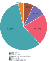
The total number of patients was 280. Baseline characteristics are shown in Table I. Out of them, 145 (51.8%) patients were males. Mean age was 47.14 ±12.8 years and 154 (55.4%) patients were more than 45 years old. The most common presenting complaint was abdominal pain, which was noticed in 212 (75.7%) patients, followed by jaundice in 204 (72.9%) patients and weight loss in 143 (51.2%) patients. The most common indication for ERCP was common bile duct (CBD) stricture, seen in 164 (58%) patients, followed by CBD stone in 60 (21%) patients and chronic pancreatitis in 26 (9%) patients (Figure 1). Bile Culture was positive in 195 (69.6%) patients and post-ERCP cholangitis developed in 187 (66.8%) patients. The most common organism in bile culture was Escherichia coli (E. coli) seen in 82 (42%) followed by Pseudomonas aeruginosa in 50 (25.6%) patients, multiple organisms in 25 (12.8%), Klebsiella in 24 (12.3%), Acinetobacter in 12 (6.1%), and Enterococcus in 2 (1%) patients (Table II).
Table I
Baseline characteristics and indications of ERCP in the studied population (n = 280)
Table II
Microbial profile of bile culture (n = 195)
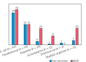
Out of 195 positive bile cultures, MDR organisms were noted in 119 (61%) patients. The most common cause of MDR infection was E. coli, seen in 43 (36.1%) patients, followed by Pseudomonas aeruginosa in 25 (21%), Klebsiella in 20 (16.8%), multiple organisms in 20 (16.8%), and Acinetobacter in 11 (9%) patients (Figure 2). Most commonly, organisms were sensitive to carbapenems followed by piperacillin/tazobactam, tigecycline, cefepime, ceftriaxone, ampicillin, amoxicillin, and ciprofloxacin, respectively.
Among the 195 patients with positive bile culture, 179 developed post-ERCP cholangitis. On multivariate analysis, preoperative jaundice, history of abdominal pain, and weight loss on admission along with ERCP performed for CBD stricture and CBD stone were independent predictors of positive bile culture on ERCP (Tables III and IV), while age greater than 45 years, presence of preoperative jaundice, abdominal pain and weight loss, positive bile culture along with ERCP performed for CBD stricture were independent risk factors for the development of post-ERCP cholangitis (Tables V and VI).
Table III
Univariate analysis for risk factors for positive bile culture
Table IV
Multivariate analysis for risk factors for positive bile culture
Table V
Univariate analysis for risk factors for post-ERCP cholangitis
Table VI
Multivariate analysis for risk factors predictive of post-ERCP cholangitis
Discussion
The sterility and continuous bile flow in the biliary system is an unfavourable medium for bacterial or organism growth. Blockage of bile duct due to any aetiology can result in bile stasis, allowing the bacteria and other organisms that are transferred through duodenal papillae to reside, replicate, and colonize in the bile duct, resulting in severe consequences including cholangitis and biliary tract infection [8].
Herein, we identified the microbial profile in the patients undergoing index ERCP. The bile culture positivity rate was 74.6%, i.e. 209 out of 279 patients had positive bile culture. Gram-negative bacteria was the leading cause of bacterial growth, accounting for 86% of the positive cultures. Among Gram-negative bacteria, E.coli was the leading cause, followed by Pseudomonas aeruginosa, multiple organsims , Acinobacter, Klebseilla, and Enterococci species. Our microbial profile was comparable to that seen in other studies, and similar to intestinal flora [8–11]. A study done by Hadi et al. [12] reported 36% positivity of bile culture in patients undergoing cholecystectomy with Gram-negative bacteria, accounting for 80% of the positive cultures, with a microbial profile similar to that of our population. Similarly, Ruan et al. reported a bile culture positivity rate of 38% in patients undergoing ERCP in a Chinese population [11]. In comparison to other studies, the high positivity of bile culture in our population was due to prolonged stasis of bile prior to ERCP, because the most common indication for ERCP in our patients was CBD stricture followed by CBD stone. There are other studies reporting variable bile culture positivity rates in different populations, ranging from 16% to 85% [9, 13–18].
In our study, bile was colonized by a single organism in most of the cases, as compared to the multiple organisms. This finding was comparable with the other studies – Ruan et al. and Kaya et al. revealed similar results [9, 11]. In contrast, a few other studies reported higher rates of multimicrobial growth in bile culture [14, 19]. This difference might be due to prolonged and overuse of antibiotics prophylactically prior to the ERCP in some areas or due to substandard culture medium.
E. coli was the most common strain colonizing the biliary tract, followed by Pseudomonas, Acinetobacter, Klebseilla, and Enterococci. E. coli is a common organism colonizing the gastrointestinal tract, while Klebseilla and Acinetobacter colonize both the gastrointestinal and respiratory tracts causing opportunistic infections [20, 21]. MDR infections were more common in our population due to easy availability of the broad spectrum antibiotics, which are prophylactically prescribed by local general practitioners. The recommended prophylactic treatment option for the prevention of PEC in patients undergoing ERCP includes cephalosporin or β-lactamase antibiotics [22]. Considering the prevalence of MDR infections in our population, most organisms were sensitive to carbapenems, followed by β-lactamase antibiotics and cephalosporins. The knowledge of this microbial profile and their antibiotic sensitivities will aid us in the usage of appropriate antibiotics for these organisms prophylactically and can also help in avoidance of antibiotic resistance.
On multivariate analysis, presence of jaundice, abdominal pain, and weight loss on admission together with ERCP performed for CBD stone and stricture were independent factors predictive of positive bile culture. This higher tendency of positive bile culture in patients with prolonged CBD stricture can be explained by the fact that biliary malignancies result in cachexia and malnutrition, leading to weight loss and decreased immunity and impaired mucosal response to pathogens resulting in bile colonization of opportunistic organisms and infection. Prolonged stasis and longer disease duration, as seen in patients with either benign or malignant CBD stricture or CBD stone, also result in biliary obstruction and proximal biliary tree dilatation causing bile stasis resulting in microbial retention and growth.
In our study, we also found that patients presenting with either jaundice, abdominal pain, weight loss with or without advanced age, undergoing ERCP for CBD stricture and positive bile culture at ERCP were at high risk of developing PEC. This can again be explained by the fact that all these findings are seen in prolonged stasis due to malignant strictures resulting in cachexia, malnutrition, and impaired immunity leading to impaired mucosal barrier causing microbial colonization and PEC. A study by Mahafzah and Daradkeh showed positive correlation between bile culture positivity and advanced age [23]. In our study, similar results were noted as the patients having age greater than 45 years were at increased risk of PEC. The knowledge of these factors causing PEC can help the clinician to start the broad-spectrum antibiotics prior to ERCP after a discussion with a multidisciplinary team involving, in particular, the infectious disease team.
Currently, prophylactic antibiotic treatment prior to ERCP is not routinely recommended. However, it is required when there is a need of repeated biliary interventions to achieve adequate biliary decompressions [22, 24].
There were certain limitations to our study. Firstly, it was a single-centred study, and secondly, the sample size was small. Hence, there is a need for a multicentric study with larger sample size to recommend prophylactic antibiotics to patients who are at high risk of developing PEC.
However, the strength of our study was that it was cross-sectional and pioneering study from this part of the world that revealed the microbial profile of biliary tract and risk factors predictive of PEC cholangitis. We followed strict protocols to perform all ERCP procedures by sterilizing all the instruments according to the international standards prior to the procedure to avoid cross-transmission between the patients.
Conclusions
The microbial profile and risk factors for positive bile culture and PEC were evaluated. Advanced age, pre-operative jaundice, abdominal pain, and weight loss along with prolonged biliary stasis were independent risk factors for these conditions. Therefore, there should be a proper preoperative management plan including a multidisciplinary approach for the commencement of prophylactic antibiotic therapy in these high-risk patients prior to ERCP.


